
Developmental Anatomy
Stage 18: Summary
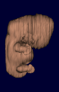
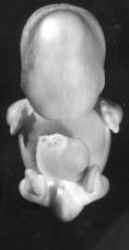
External: The head is relatively larger than previously; the main axis of the trunk has become straighter, and a slight indication of a lumbar curvature may be found; the nasal pit is further medial and is directed ventrally so that the nostril is not visible in profile views, and nasofrontal grooves are distinct; the full complement of auricular hillocks is present on the mandibular and hyoid arches; the hand plate exhibits definite digital rays, and the foot has acquired a rounded digital plate; surface elevations of individual somites are becoming limited mostly to the lumbosacral region.
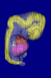
Internal: In the heart, foramen secundum and semi-lunar cusps have appeared (stages 15-17), and fora-men primum is being obliterated and the atrioventricular cushions are fusing (stages 16-18); the beginnings of the palate are appearing, the dorsal and ventral pancreas are fused, and the vermiform appendix is distinguishable; the bronchial tree shows segmental buds; the mesonephros is functional, calices are developing, and the urogenital sinus presents two divisions; chondrification begins in some of the vertebral centra and in the humerus and radius; the future olfactory bulb is indicated; the retinal fissure is largely closed and the lens cavity is becoming crescentic; semicircular ducts are imminent but none is yet present; the auditory ossicles are defined.
Lab Manual - Horizon 18 (CC#7707)
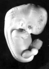
- The body of the embryo is a more unified cuboidal mass than in earlier groups, and there is some indication of both lumbar and cervical flexures.
- The extremities are somewhat longer; the digital rays of the hand are definitely notched.
- The elbow region is usually discernable, and the foot plate and toe rays can be seen in some specimens.
- Eyelid folds are present in older specimens, and the pigmented retina is partly covered by white opaque condensed scleral masses, particularly above and on the lateral side of the retina.
- A distinct tip of the nose can be seen in profile.
- Auricular hillocks are being transformed into definite parts of the external ear.
- Ovulation age: 44-48 days.

Developmental Stages in Human Embryos by Ronan O'Rahilly and Fabiola Müller.
Published by Carnegie Institution of Washington, Publication 637. 1987.
Stage 18 (PDF 1.3 MB)
QUESTION: What is the overall shape of the embryo at this point?