
3-D Modeling Valuable Tool in Forensic Discovery
By: Paul Bello, National Museum of Health and Medicine
SILVER SPRING, Md.: The National Museum of Health and Medicine (NMHM) continued its "Medical Museum Science Café" series May 27, as guests embarked on the process involved with recreating the face of an individual through a combination of art, forensic science and anthropology.
The discussion, entitled "The Science and Art of Forensic Facial Reconstruction" was led by Shayne Kondor, senior medical modeling engineer and consultant to the Naval Postgraduate Dental School in Bethesda, Md. He explained how 3-D modeling of craniofacial reproductions is done and illustrated both the classic approach that includes a clay sculpture over a skull to the more cutting-edge digital approach that involves an optical or laser surface scan.
It was through this latter technique, explained Kondor, that a forensic artist and biomedical engineer used computed tomography (CT) data of a skull first discovered in 1876 to reveal the face of an unknown Civil War soldier from the 54th Massachusetts Volunteers. That skull now sits on display at the museum.
"It takes approximately four weeks to reconstruct the face of a skull using this classic clay sculpture approach. You need to be a highly trained forensic artist and sculptor to do this," Kondor said. "The digital approach allows you to design the face on top of that model so we can do variations on that [the digital surface model of the skull]. That allows us to change features or go back and look for more evidence to help in the process of putting a face to these remains."
Through these methods, Kondor noted that identifying the age, sex and even the ancestry of a skull greatly enhance the reconstruction process. An already evolving method has gained even more traction in recent years with the introduction of cone beam (CBCT) scanners. According to Kondor, these scanners have been developed to deliver scale models at a fraction of the radiation exposure and cost associated with a conventional CT.
"It's important to have a comparison between a CBCT and conventional CT. That's so we can properly assess the viability of this technology as a means of obtaining pre-deployment records of craniofacial structures of our service members," Kondor said. "Based on certain markers it appears that three-dimensional digital models based on a CBCT scan will register to models from conventional CT scans. More importantly, the effects of CBCT exposure, its detector type and resolution also don't cause significant differences in the quality of model registration."
In addition to his work at the Naval Postgraduate Dental School, Kondor was a member of the research faculty at Georgia Tech and the Georgia Tech Research Institute. His research activities, at both institutions, included patient specific medical modeling and applications of additive manufacturing in medicine and dentistry.
Additive manufacturing is the process of joining materials to make objects from 3-D model data, usually layer upon layer, as opposed to subtractive methodologies, Kondor said. Through a computer-aided design (CAD), he noted that additive manufacturing equipment can read data from a CAD file and place layers of liquid, powder, or some type of sheet material to help fabricate a 3-D object.
NMHM has several 3-D printed objects on display in an exhibit on traumatic brain injury and others in an exhibit on advances in military medicine. Representative examples of the technology are also in the holdings of the museum's historical collection.
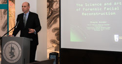 |
Caption: Shayne Kondor, a senior medical modeling engineer and consultant to the Naval Postgraduate Dental School, was guest speaker during the Medical Museum Science Café May 27, 2014 at the National Museum of Health and Medicine (NMHM) in Silver Spring, Md. The discussion was entitled: The Science and Art of Forensic Facial Reconstruction. |
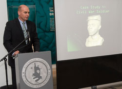 |
Caption: Shayne Kondor, a senior medical modeling engineer and consultant to the Naval Postgraduate Dental School, explains how 3-D modeling and forensic anthropology came together to reveal the face of an unknown Civil War soldier. A skull of a soldier of the revered 54th Massachusetts Volunteers is on display at the National Museum of Health and Medicine (NMHM) in an exhibit in Civil War medicine. Kondor was guest speaker during NMHM’s Medical Museum Science Café May 27, 2014. NMHM is located in Silver Spring, Md. |
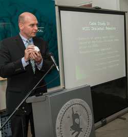 |
Caption: Shayne Kondor, a senior medical modeling engineer and consultant to the Naval Postgraduate Dental School, discusses advances in forensic anthropology while holding a small model of a human head that was first designed using 3-D modeling and computer imaging. Kondor was guest speaker during the National Museum of Health and Medicine’s (NMHM) Medical Museum Science Café held at NMHM in Silver Spring, Md. May 27, 2014. |
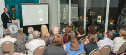 |
Caption: Medical Museum Café attendees listen to Shayne Kondor, a senior medical modeling engineer and consultant to the Naval Postgraduate Dental School (standing, far left), as he explains the inner workings of forensic anthropology and 3-D modeling. The program was held at the National Museum of Health and Medicine (NMHM) in Silver Spring, Md. May 27, 2014. |
(Disclosure: These images have been cropped to emphasize the subject.) (National Museum of Health and Medicine photo / Released) |
|