Medical Illustrators Get Under the Skin of Athletes at 2019 Anatomy of Sports
By Jacqueline Gase
NMHM Public Affairs Coordinator
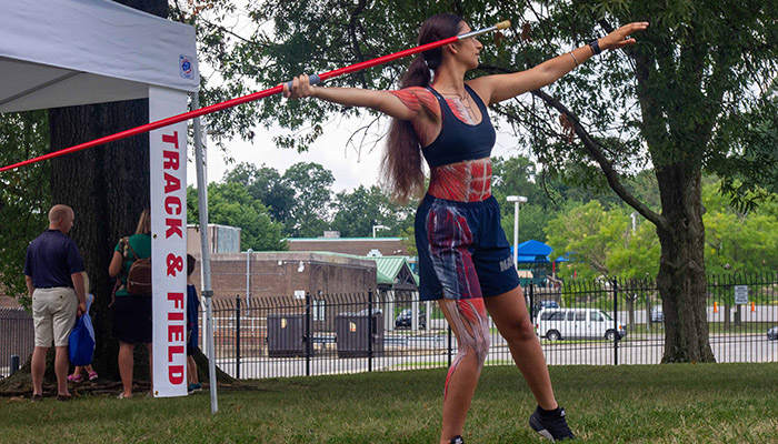
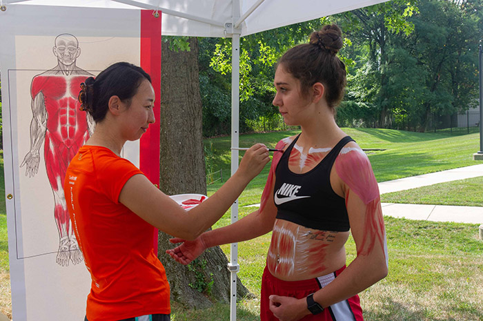
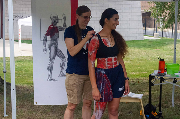
U.S. Navy Ensign Ashna Manhas prepares to throw a javelin as she shows her painted muscle anatomy during the Aug. 17, 2019, Anatomy of Sports program at the museum in Silver Spring, Maryland. (NMHM photo)
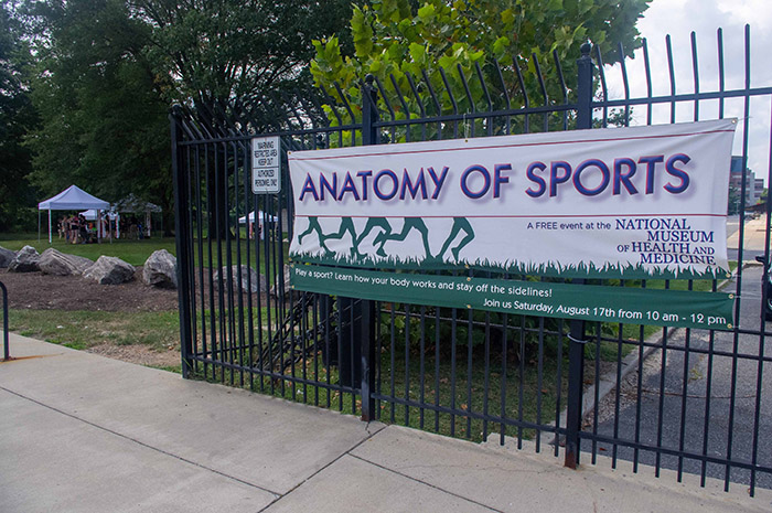
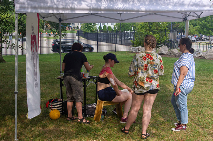
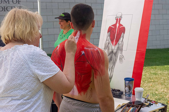
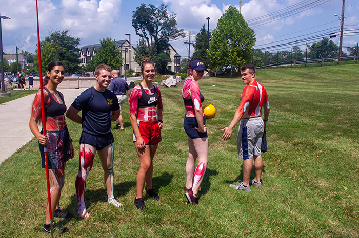
Medical illustrators, athletes, and physical therapists worked together to educate museum visitors on the anatomy of the human body at the August 17, 2019, Anatomy of Sports program at the National Museum of Health and Medicine. Medical illustrators painted the underlying skeletal muscles of Uniformed Services University of the Health Sciences (USU) athletes highlighting particular muscle groups used most frequently in the sports of football, javelin, cycling, swimming, volleyball, and horseback riding. Alongside the artists, University of Maryland (UMD) physical therapists and students and USU medical students discussed the importance of strengthening muscles to prevent injury.
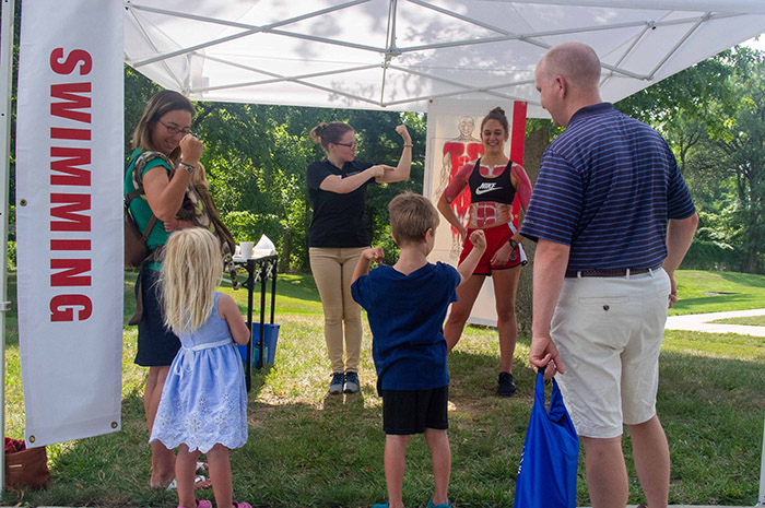
UMD physical therapist Jason Shipley commended artist Ikumi Kayama on her attention to the back muscles she painted on swimmer U.S. Army 2nd Lt. Allison Watkins. While shoulder muscles are crucial for swimming, Shipley noted that back muscles, like the latissimus dorsi, are equally important. "Regular strengthening programs for all back muscles can help prevent common swimming injuries, like ‘swimmer's shoulder,'" said Shipley.
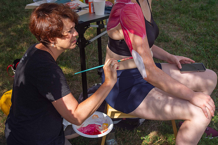
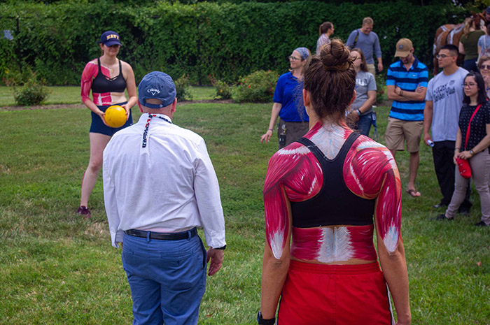
Similarly, volleyball players, like U.S. Navy Ensign Laurel Spack, must attend to the back and arms for their sport. Medical illustrator Alexandra Webber highlighted this by painting the latissimus dorsi, trapezius, deltoid, triceps, biceps, and pectorialis muscles.
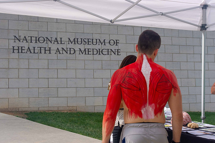
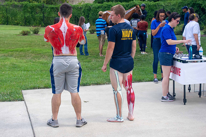
Football player U.S. Air Force 2nd Lt. Galen Gist noted that his sport consists of forceful pushing. To combat this, some football players participate in yoga. "If you're not stiff, it's harder to get injured," said Gist. Medical illustrator Marie Dauenheimer stated that ballet is also a training exercise that helps strengthen back muscles similar to the ones she painted on Gist.
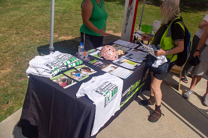
Stationed near Gist and Dauenheimer, a presenter from the Defense and Veterans Brain Injury Center's "A Head for the Future" initiative informed museum visitors about preventing, recognizing, and recovering from a common injury in football: traumatic brain injury.
At another tent, USUHS medical illustrator Elizabeth Weissbrod, paid special attention to the abdominal and shoulder muscles while painting javelin thrower U.S. Navy Ensign Ashna Manhas. Manhas explained that the twisting of her body when throwing the javelin relies heavily upon the external obliques of her abdomen. UMD physical therapy student Karson Becker also noted that a rotator cuff muscle, the supraspinatus, is a key muscle for the sport.
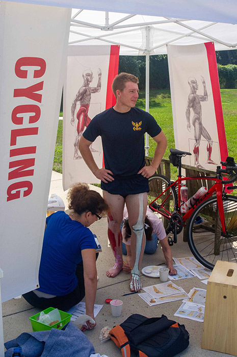
Conversely, the hamstrings, gastrocnemius, and quadriceps femoris are all deemed critical by cyclist U.S. Navy Ensign Zech Brooke. Medical illustrators Joan Tycko and Alice Tangerini actualized the muscles and bones while UMD physical therapy student Nicole McKenzie discussed how cycling is a recommended therapy for people with knee injuries.
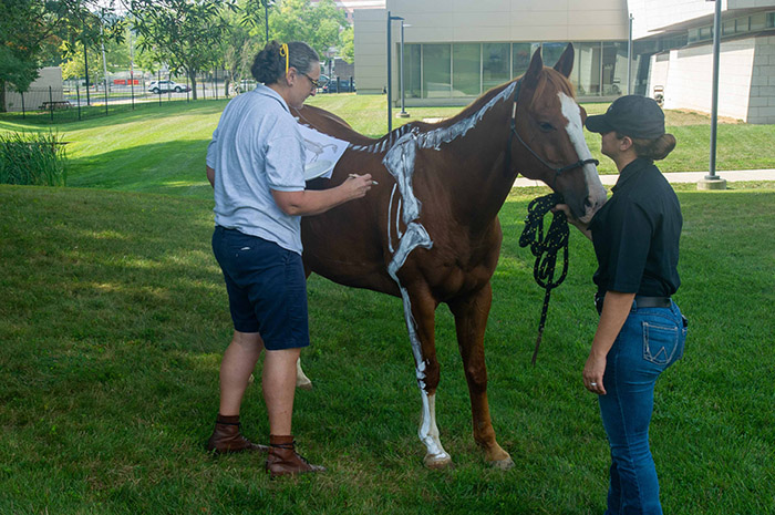
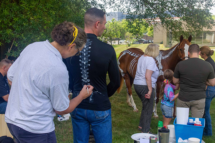
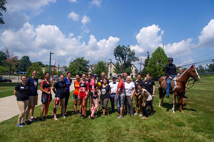
Museum visitors were also treated to animal athlete Skeeter, a therapy horse from the Fort Myer Caisson Platoon. The museum's Human Developmental Anatomy Center Collections Manager and medical illustrator Elizabeth Lockett painted the skeletal anatomy of both Skeeter and his rider U.S. Army Sgt. 1st Class Michael Skeen. UMD physical therapist Vincent Conroy noted the similarities in anatomy, saying "we [humans and horses] rely on all the segments of our vertebrae to work. If we restrict one segment it limits the ability to move."
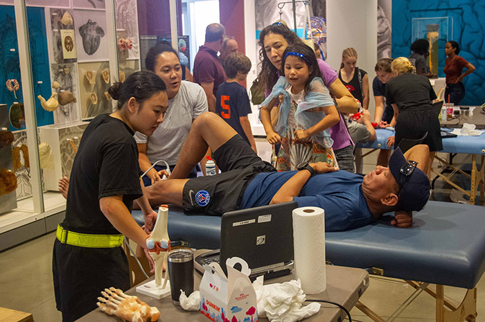
Inside the museum, visitors saw beneath the skin with technology. Using an ultrasound machine, USU students showed visitors their muscles, veins, and bones.
"While the program is fun and illustrates what the human eye can't see, it's also instructive because it reminds visitors to pay special attention to strengthening their muscles so they don't get hurt," said Andrea Schierkolk, the museum's public programs manager. "This is especially important for our service members since medical readiness is vital for them to stay safe when undertaking challenging physical activities," said Schierkolk.
The program also highlights the value of medical illustration to record and disseminate anatomical and medical knowledge. The museum's Otis Historical Archives Medical Illustrations Collection preserves medical art ranging from the nineteenth century to World War II.
The museum's public programs provide forums for informal learning that connect the mission of the Department of Defense museum with the public. For more information about upcoming events, call (301) 319-3303 or visit https://www.medicalmuseum.mil.