
A Fragile Organ: The Brain
Anatomy and Pathology
Anatomy is the study of the body, a complex system of interrelated structures that work together to maintain health. Sometimes these structures break down. By studying anatomical specimens, researchers investigate how injuries and disease affect the body. Anatomy and pathology, combined with advanced imaging technologies, improve our understanding of how the body works.
A Fragile Organ
U.S. Army Maj. Walter Reed (1851-1902) (NCP 0597)
The brain is a delicate and intricate structure. Each of our bodily functions—from basic processes like breathing to the highly complex systems that control speech or reasoning—is regulated by a different part of the brain. The brain acts as the body's overall decision maker, coordinating the continuous exchange of voluntary and involuntary responses that guide our every action. Although the brain is protected by a cushioning layer of fluid surrounded by the thick bones of the human skull, injury and disease can still have a devastating impact. Learning how the brain works helps researchers explore new treatments and cures.
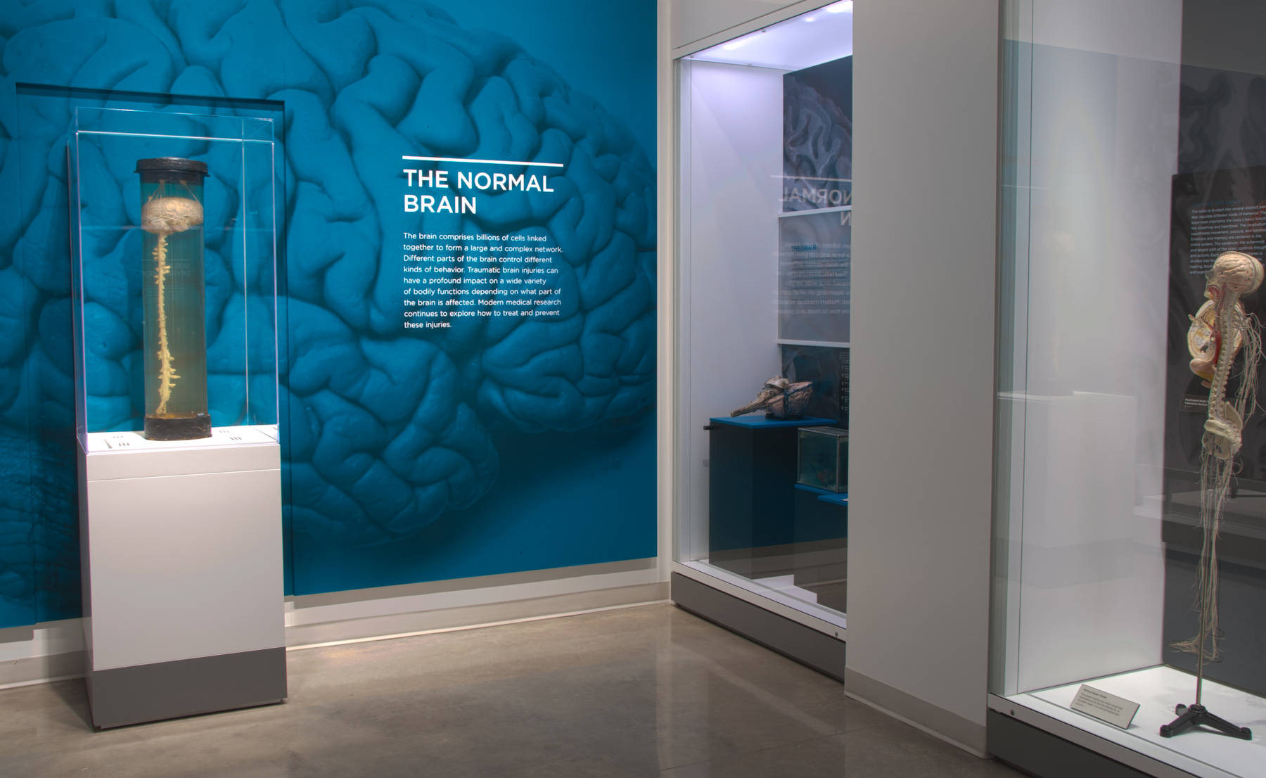
The Normal Brain
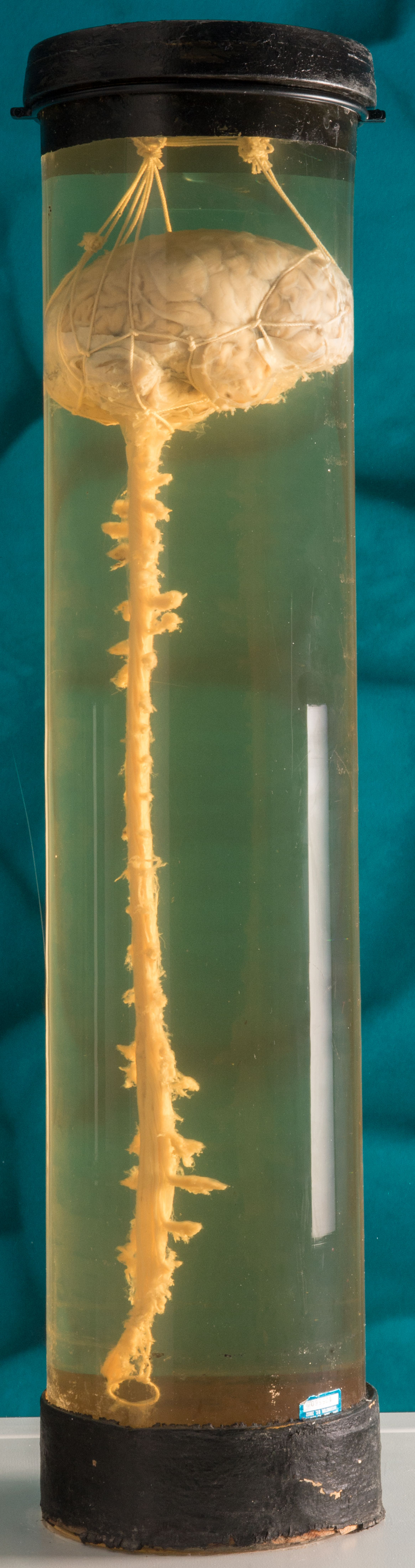
Wet Brain and Spinal Cord, 1935. The spinal cord is a complex bundle of axons, or nerve fibers, that carry information from the brain to the torso, arms, and legs. (NMHM 1987.0003.27) (National Museum of Health and Medicine photo by Matthew Breitbart / Released)
The brain comprises billions of cells linked together to form a large and complex network. Different parts of the brain control different kinds of behavior. Traumatic brain injuries can have a profound impact on a wide variety of bodily functions depending on what part of the brain is affected. Modern medical research continues to explore the best ways to treat and prevent these injuries.
Traumatic Brain Injury
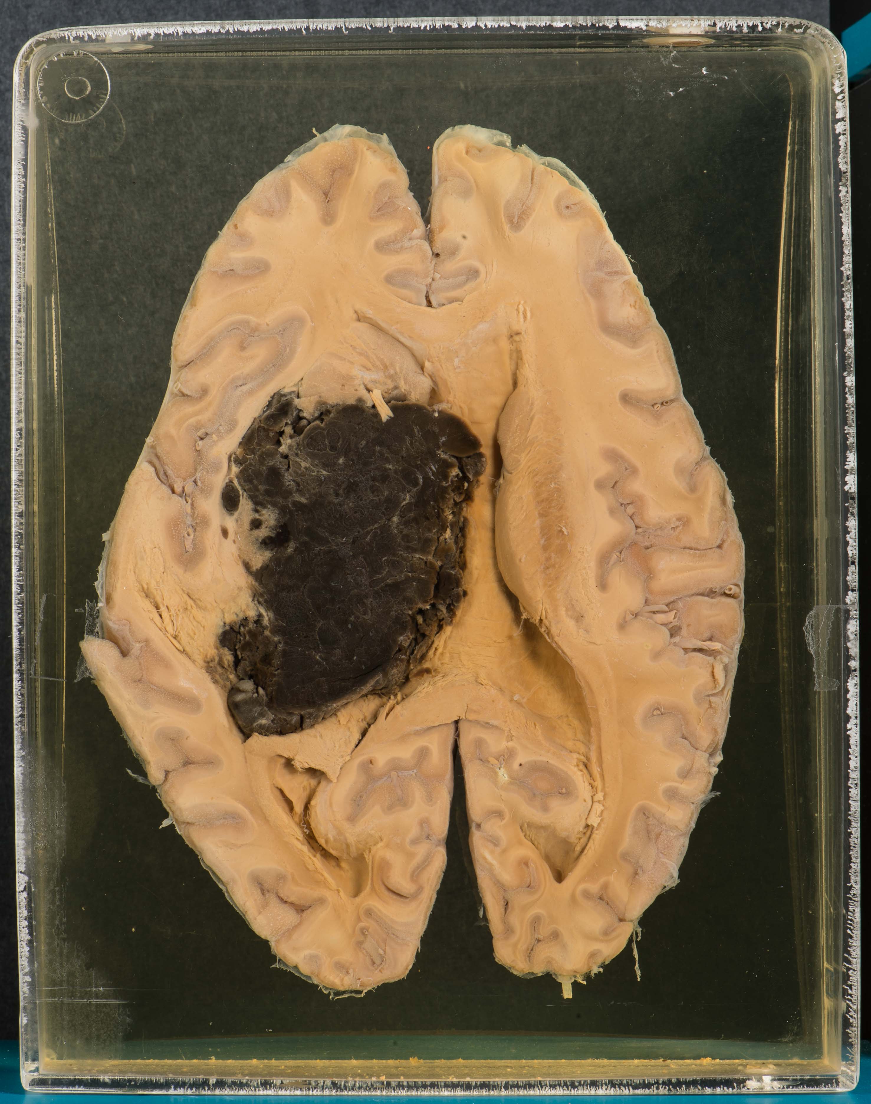
Brain, Intracerebral Hemorrhage. (AFIP 0001917) (National Museum of Health and Medicine photo by Matthew Breitbart / Released)
Traumatic brain injury (TBI) occurs when the brain is damaged by a sudden force. If the damage affects the parts of the brain that control these functions, TBI can have a drastic impact on mental abilities, personality, and memory. Although millions of civilians suffer from TBI, military service members are especially vulnerable. Explosive devices are a leading cause of TBI for active duty personnel in war zones, and TBI has become one of the signature injuries of modern warfare.
Diagnosing TBI
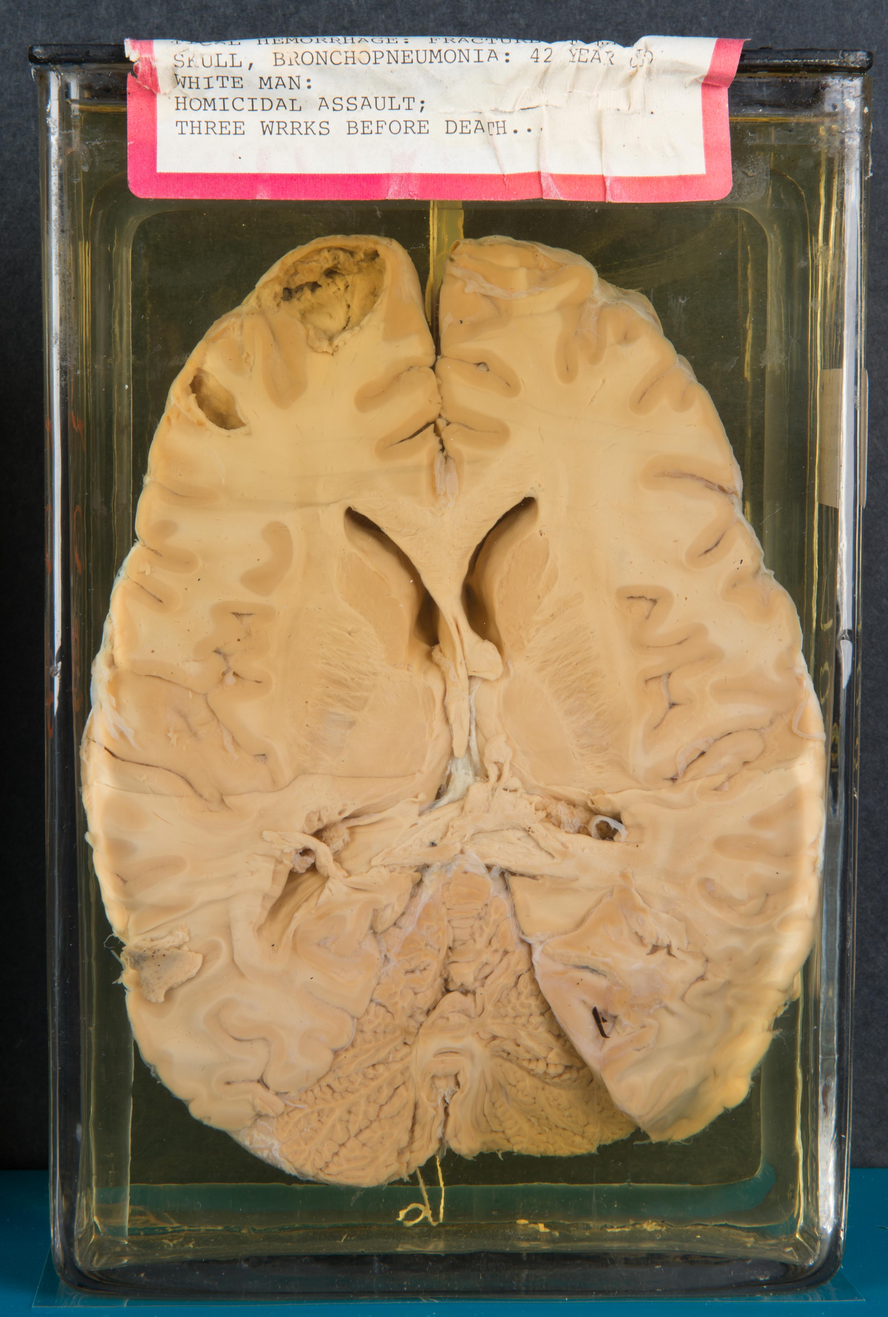
Brain, Extensive Coup-Contrecoup Lacerations with Subcortical Hemorrhage. A coup-contrecoup injury occurs when an impact or violent motion brings the head to a sudden stop, causing the brain to slam into the skull. The brain is injured at the point of impact, and because it bounces back into the opposite side of the skull, the opposite side of the brain also is injured. (1990.0003.1237) (National Museum of Health and Medicine photo by Matthew Breitbart / Released)
Diagnosing TBI can be difficult. Doctors and medics look for signs, such as unconsciousness, memory loss, or decreased ability to understand and respond to commands. Medical personnel often also use the Glasgow Coma Scale (GCS), a neurological test that measures a patient's eye, verbal, and motor responses. The GCS ranges from 3 (a deep coma) to 15 (normal behavior). The lower the score, the more likely the patient suffers from TBI.
Treating TBI
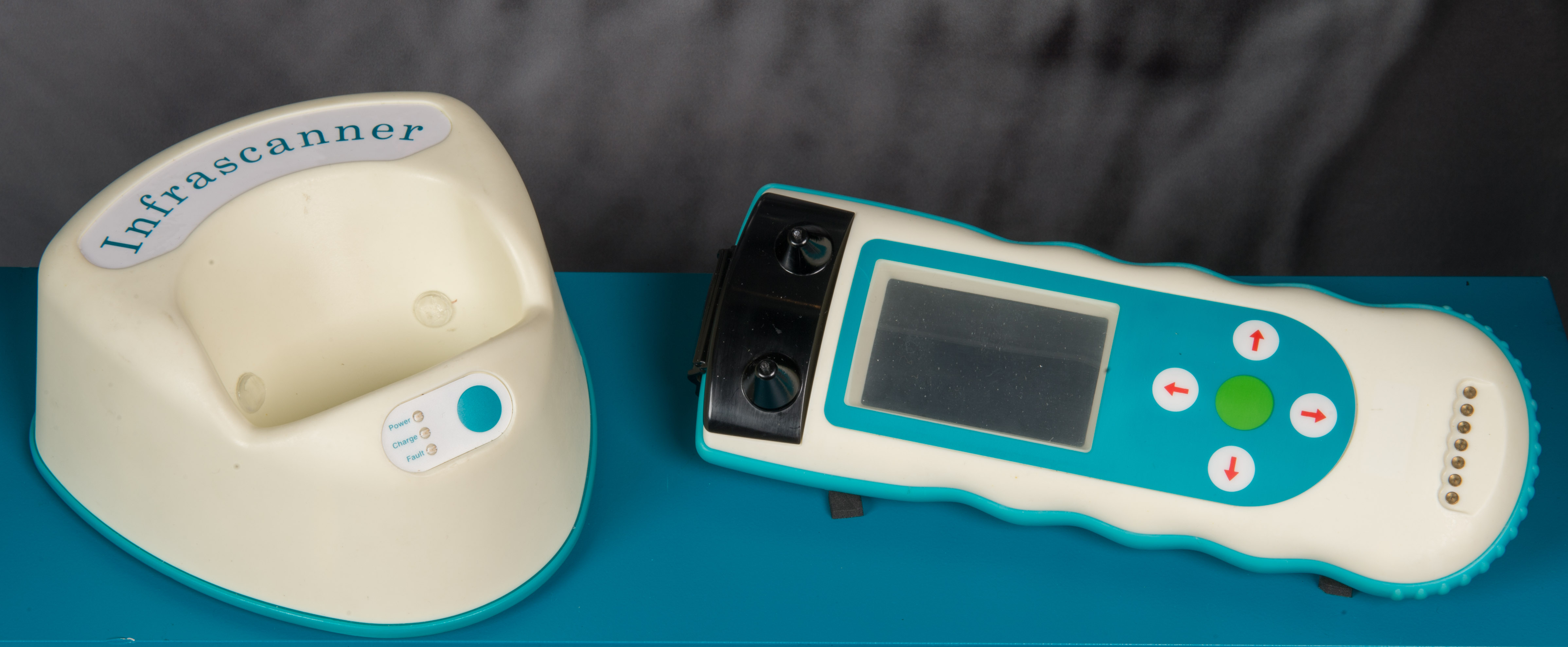
Near Infrared (NIR) system for detection of brain hematomas, invented by Dr. Britton Chance (University of Pennsylvania) and Dr. Claudia Robertson (Baylor College of Medicine) and manufactured by InfraScan, Inc. The device is a notable advancement in combat casualty care as a triage tool for one of the most problematic diagnoses on the battlefield: traumatic brain injury with intracranial bleeding. The lack of CT-scanning technology in the field can result in this diagnosis being missed or mistreated. This second-generation Infrascanner Model 2000 is an updated unit with the wireless capabilities built into the device, eliminating the need for an external PDA and is more "rugged" to withstand rain, dust, salt, vibrations, and being dropped. Its accessories include a disposable black plastic scanning shield, cradle/charging stand, and power supply cord. The 2000 model was tested in Afghanistan from 2011-2012. The field-studies conducted by Marines proved it to be a useful tool for the military. It was FDA-approved in January 2013. (National Museum of Health and Medicine photo by Matthew Breitbart/ Released)
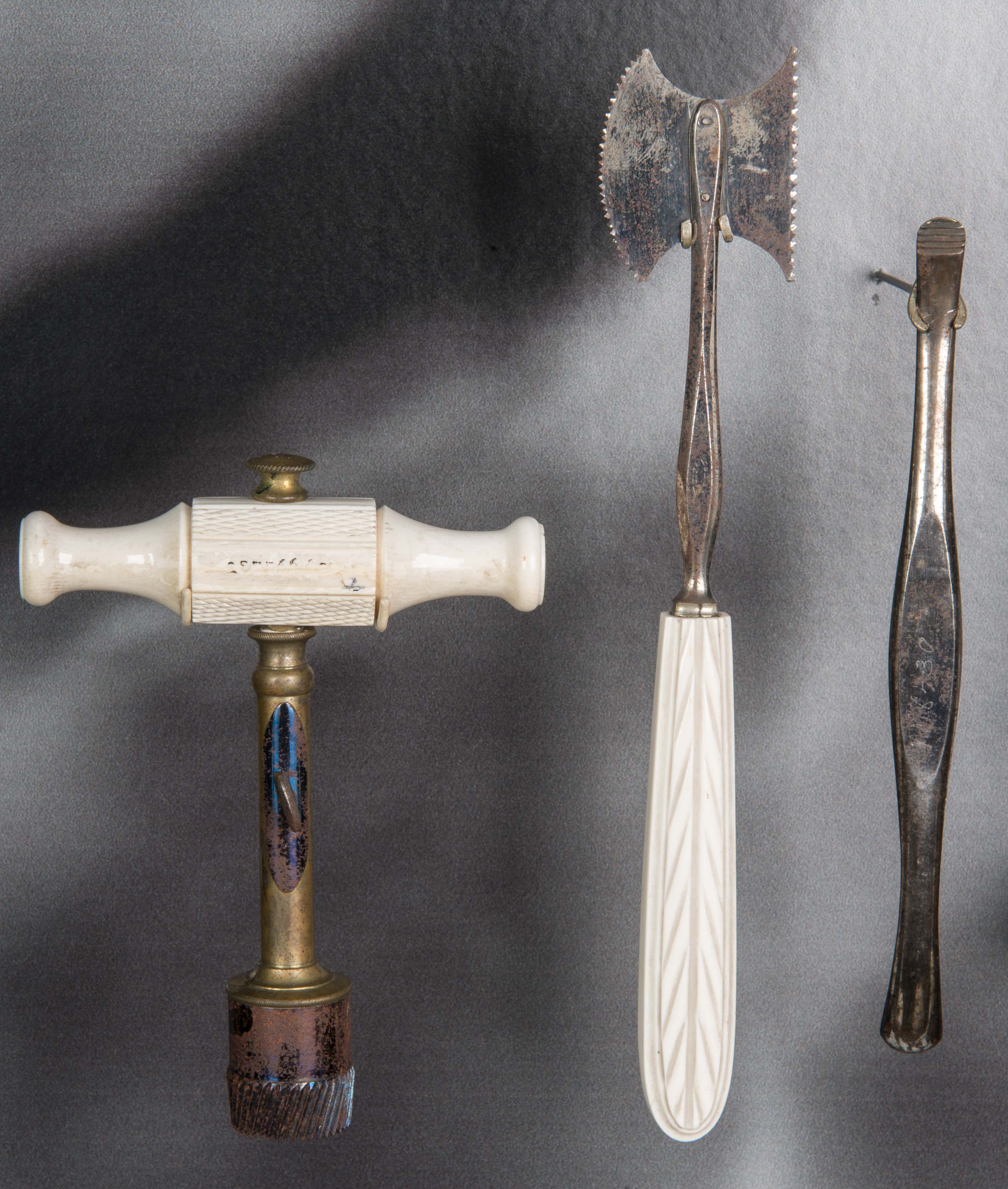
Trephination Tools that Belonged to Surgeon General William Hammond, ca. 1860. (M-129.00088) (National Museum of Health and Medicine photo by Matthew Breitbart/ Released)
An injured brain tends to swell, which can cause additional damage and lead to life-threatening blood flow problems. One option for relieving high intracranial pressure is to create a small opening in the patient's skull. Physicians have been performing this surgery for thousands of years using a variety of tools and devices. Today, neurosurgeons use modern instruments to perform craniotomies—a surgical procedure to alleviate pressure by removing a bone flap from the skull.