
Discovery of the X-ray: A New Kind of Invisible Light
November 8 is World Radiography Day, the anniversary of Wilhelm Röentgen's discovery of "a new kind of invisible light" -- the X-ray.
Röentgen discovered X-rays accidentally while doing experiments on fluorescence produced in vacuum tubes. Working with a completely shielded Crookes cathode tube, he noticed that rays emitted from the tube somehow passed through the shielding, making a shadow-like image on a light-sensitive paper nearby. Following up on this observation, on November 8, 1895, Röentgen produced an image of the bones of his wife's hand as evidence of his discovery.
X-ray technology revolutionized medicine by providing a way to view interior structures of the human body without invasive or exploratory surgical procedures, giving new insights into injury and disease, and allowing for thoughtful planning before treatment.
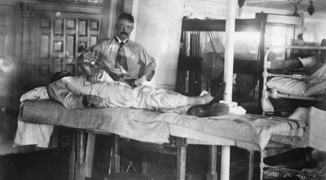 No. 89. Taking an X-ray photograph on board U.S.A. Hospital Ship Relief, 1898 (OHA313-089)
Röntgen's discovery saw immediate and widespread integration in the medical field. In this photograph, Dr. William Gray of the Army Medical Museum uses an unshielded X-ray tube above a patient's chest aboard the USAHS Relief off the coast of Cuba in 1898. Electricity for the tube is produced by the electrostatic generator situated behind the mast. On July 30, 1898, The New York Times could report, "In light of our recent experience the X ray has become an indispensable diagnostic resource to the military surgeon in active service."
Röntgen and the Discovery

Bronze bust, Christabel Cummings, 1940
Wilhelm Conrad Röntgen was born on March 27, 1845 in Germany. His family moved to the Netherlands when he was just three, and Röntgen grew up to study mechanical engineering and later physics in university. He earned his Ph.D. at the University of Zurich in 1869 and went on to become a Lecturer and Professor at various european universities. His discovery of the X-ray occurred on November 8, 1895, propelling his career even farther, and in 1900, Röntgen settled into the role of Chair of Physics at the University of Munich where he remained the rest of his career. He was awarded the Nobel Prize in Physics in 1901. (M- 360.10059)
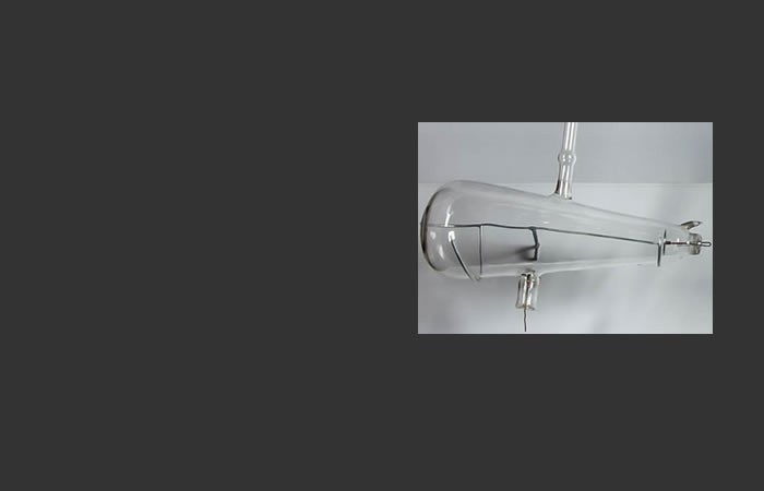
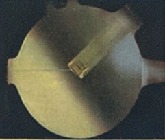
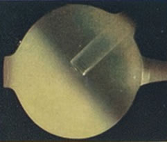
Crookes tube, named for English physicist William Crookes, is a generic name for a family of early sealed glass vessels with an electrode at either end, used to study the travel of electrical charges through a vacuum or gas and the resulting fluorescence. Johann Hittorf was the first to realize, in 1869, that most of the electrical charge within the tube traveled in a straight line between the cathode and anode, thus the name cathode "rays." This tube, with its broad-based bottle shape, is thought to be the type Röntgen was using when he discovered that besides visible fluorescence, a Crookes cathode tube also produced an invisible form of radiation, subsequently named "X-rays." (M-736.50406)
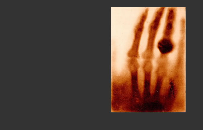
While experimenting with cathode radiation, Röntgen discovered that the rays passed easily through the flesh while shadows were cast by the bones and the ring of his wife's hand. Upon looking at the image of her skeleton, Ludwig exclaimed, "I have seen my death!" (A030067, National Library of Medicine)
"Now professor, will you tell me the history of the discovery?"
"There is no history," Röntgen replied. "I have been for a long time interested in the problem of the cathode rays from a vacuum tube, as studied by Hertz and Lenard. I had followed theirs, and other researches, with great interest, and determined as soon as I had time, to make some researches of my own. This time I found at the close of last October. I had been at work for some days when I discovered something new."
"What was the date?"
"The eighth of November."
"And what was the discovery?"
"I was working with a Crookes tube covered by a shield of black cardboard. A piece of barium platino-cyanide paper lay on the bench there. I had been passing a current through the tube, and I noticed a peculiar black line across the paper."
"What of it?"
"The effect was one of which could only be produced, in ordinary parlance, by the passage of light. No light could come from the tube, because the shield which covered it was impervious to any light known, even that of the electric arc."
"And what did you think?"
"I did not think; I investigated. I assumed that the effect must have come from the tube, since its character indicated that it could come from nowhere else. I tested it. In a few minutes there was no doubt about it. Rays were coming from the tube which had a luminous effect upon the paper. I tried it at greater and greater distances, even at two metres [sic]. It seemed at first a new kind of invisible light. It was clearly something new, something unrecorded."
"Is it light?"
"No."
"Is it electricity?"
"Not in any known form."
"What is it?"
"I don't know."
- Interview with Wilhelm Röntgen published in McClure's Magazine, April 1896.
Production of X-rays
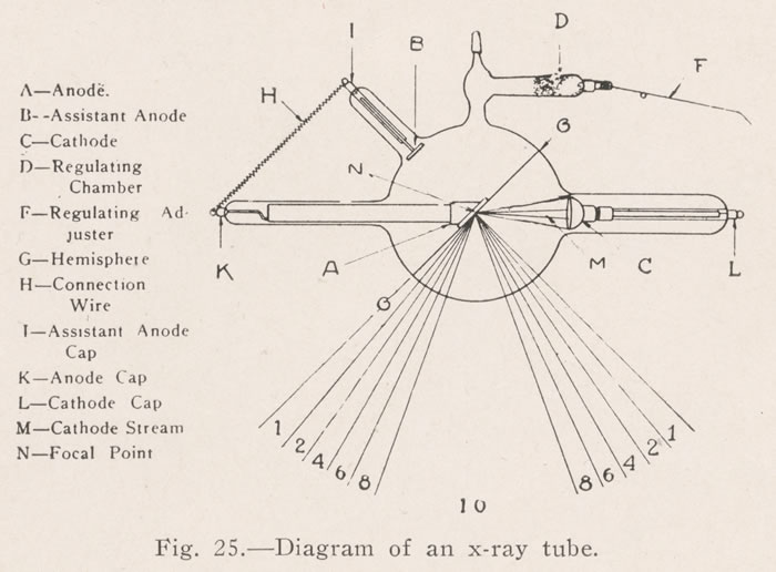 Diagram of an X-ray tube from Dental and Oral Radiography: A Textbook for Students
and Practitioners of Dentistry, James D. McCoy, 1919; NMHM Library
Diagram of an X-ray tube from Dental and Oral Radiography: A Textbook for Students
and Practitioners of Dentistry, James D. McCoy, 1919; NMHM Library
X-rays are produced by electron excitation, and are shorter in wavelength than both visible and ultraviolet light. When a current is applied to the cathode (C), electrons become agitated and escape from their atoms to form an electron cloud. In the X-ray tube, most of the electrons from this cloud will travel straight across the near-vacuum inside the tube to the anode (A), which also has an electric charge. This causes a stream of electrons to strike the target (the angled plate at center of tube), dislocating atoms of the target and producing a focused stream of X-ray radiation.
The different sizes and shapes of X-ray tubes direct the X-rays in different, useful directions. The target can be shaped and tilted to direct the rays in a narrow or broad range. Medical X-ray tubes are generally designed to facilitate the production of radiographic images. Photographic film is placed behind a patient and focused X-rays projected through the body. The differences in density of bones, flesh, organs, blood vessels, and foreign objects, absorb or interrupt the X-rays to different degrees, creating the "shadow" of these elements on the film.
Soft and Hard X-ray Tubes
The terms "hard" and "soft" were applied to X-ray tubes to describe the degree of vacuum inside the tube.
A "soft" tube had a vacuum inside that was high and generated X-rays unable to penetrate thick or dense material. A tube in this condition would be used for areas like the knees, shoulders, and elbows. The radiologist would operate the tube using short exposure times and lower electrical currents.
Images from Radiography and Radio-Therapeutics, Part I, Radiography, Robert Knox, 1923; NMHM Library
As the X-ray tube was used, the vacuum decreased, making it a "hard" tube. The resulting X-rays had greater penetration and could be used to radiograph the kidneys, spine, or skull. At some point, however, the tube becomes too "hard," and at this point it can't be regulated for standard exposures. To soften a hard tube, it can be baked in an oven or placed in a warm area until the gas inside is expelled and the vacuum is returned.
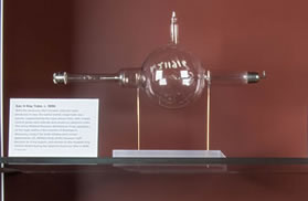
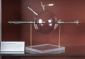
X-ray Tubes in the NMHM Historical Collection


X-ray Images
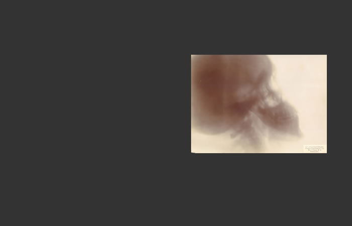
This image of the skull taken with an early gas X-ray tube is less crisp than the skull image to the right, taken in 1941. Tissues of varying density are less clearly distinguished. (CP 3614B)
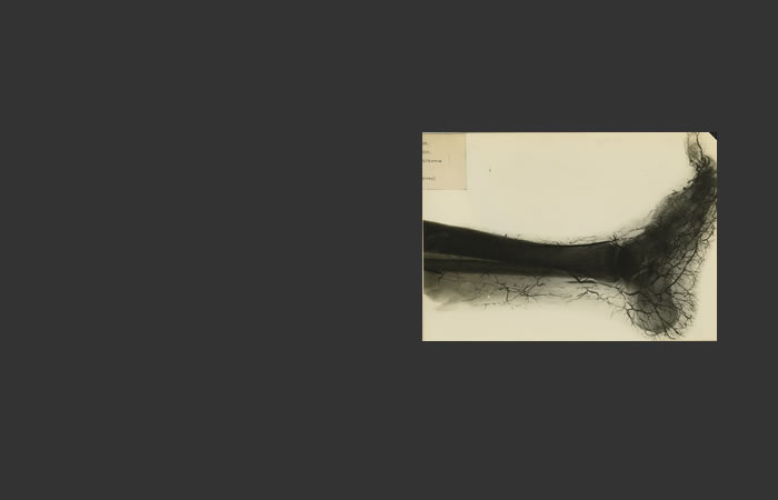
Buerger disease is caused by small blood vessels that become inflamed and swollen. This X-ray image was likely made with the improved Coolidge hot-cathode tube developed in 1913. (Reeve 043536)

This image of the lateral view of a skull shows greater detail between the various areas of bone and tissue, as well as a greater clarity of lines, compared to the skull radiographed in 1897. (The sinus and nasal areas are obstructed in this view, and those areas were radiographed separately.)
War Department Technical Manual, TM 8-240, Röntgenographic Technicians, July 3, 1941 (NMHM Library)