About The Micrograph
Welcome to The Micrograph, where you can get a glimpse behind-the-scenes at the National Museum of Health and Medicine (NMHM).
: a graphic reproduction of the image of an object formed by a microscope
The name, The Micrograph, is a metaphor for the close-up snapshot of the museum and related topics that you'll find here. It also references NMHM's microscope collection of more than 1,700 items, and a collection of photomicrographs that document the pioneering work in magnified photographic imaging by Assistant Surgeon General Joseph J. Woodward.
Techniques of photomicrography improved greatly from the early 1800s to the advent of electron microscopy in the 1930s and modern-day digital imaging. Similarly, the topics explored on The Micrograph span from historical antecedents to contemporary innovations.
In support of the NMHM mission, The Micrograph seeks to educate and inspire readers on a range of topics in health and medicine, while fostering an appreciation for the role of military medicine and its close relationship with civilian medicine.
Launched in March 2019, The Micrograph is a digital project of the NMHM community that features stories told by collections staff, researchers, educators, volunteers, staff writers, and others.
Join us as we explore the happenings and holdings at NMHM.
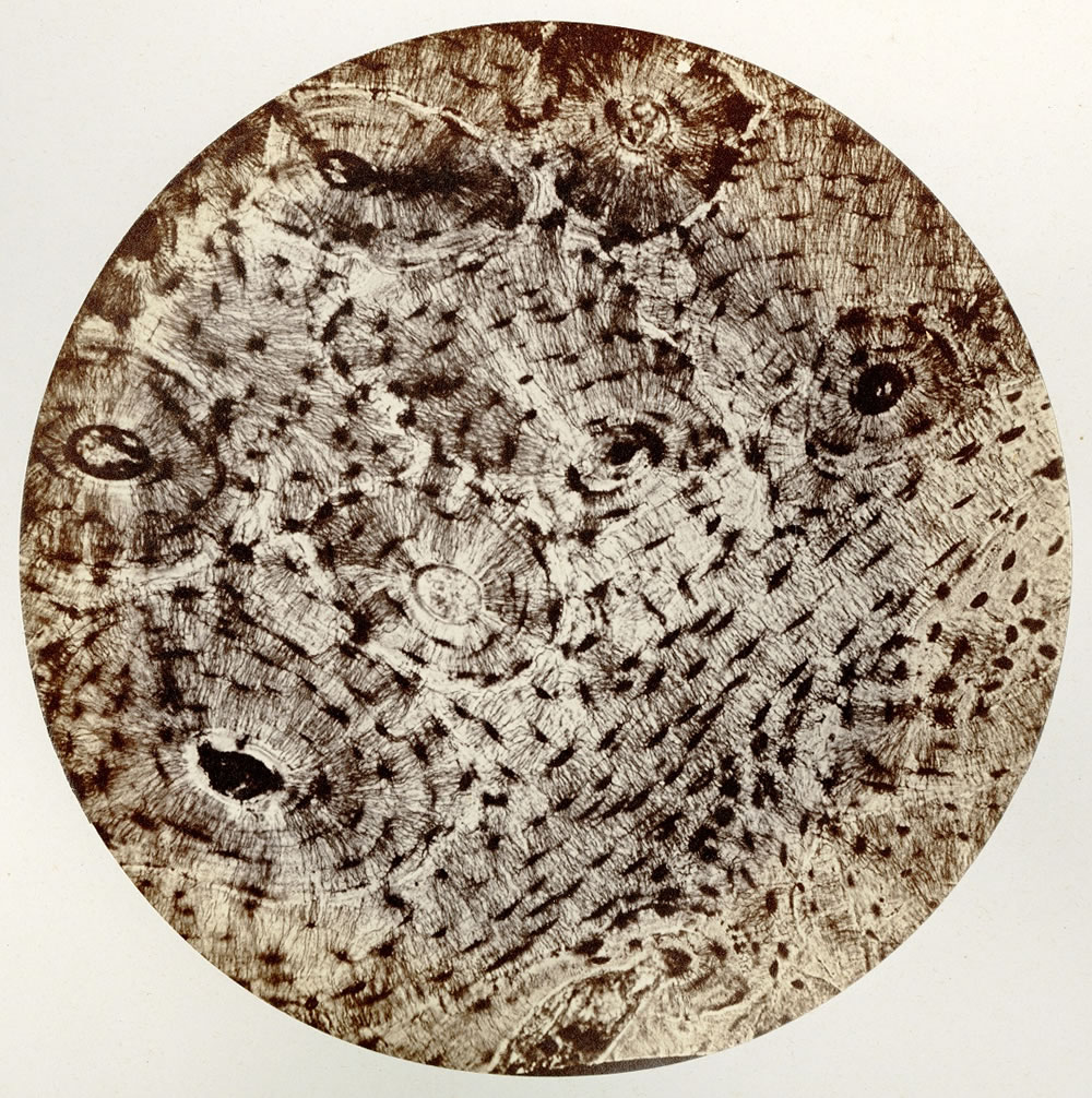
The background image used for The Micrograph shows the cross-section of a femur bone as captured in a photomicrograph made by J. J. Woodward. Click on the story to the right to learn more about the history of photomicrography and its ties to the history of the museum. (NMHM, OHA 79, No. 126)
Be sure to check out available resources and find more information about NMHM's collections,
exhibits, educational resources, and
upcoming events. Commenting is not a supported function, but we would love to hear from you.
Please email us here with any feedback you have, and use
these social media icons  found on each story page to share things that interest you on your platform of choice.

 An official website of the United States government
 ) or https:// means you've safely connected to the .mil website. Share sensitive information only on official, secure websites.

