An official website of the United States government
 ) or https:// means you've safely connected to the .mil website. Share sensitive information only on official, secure websites.


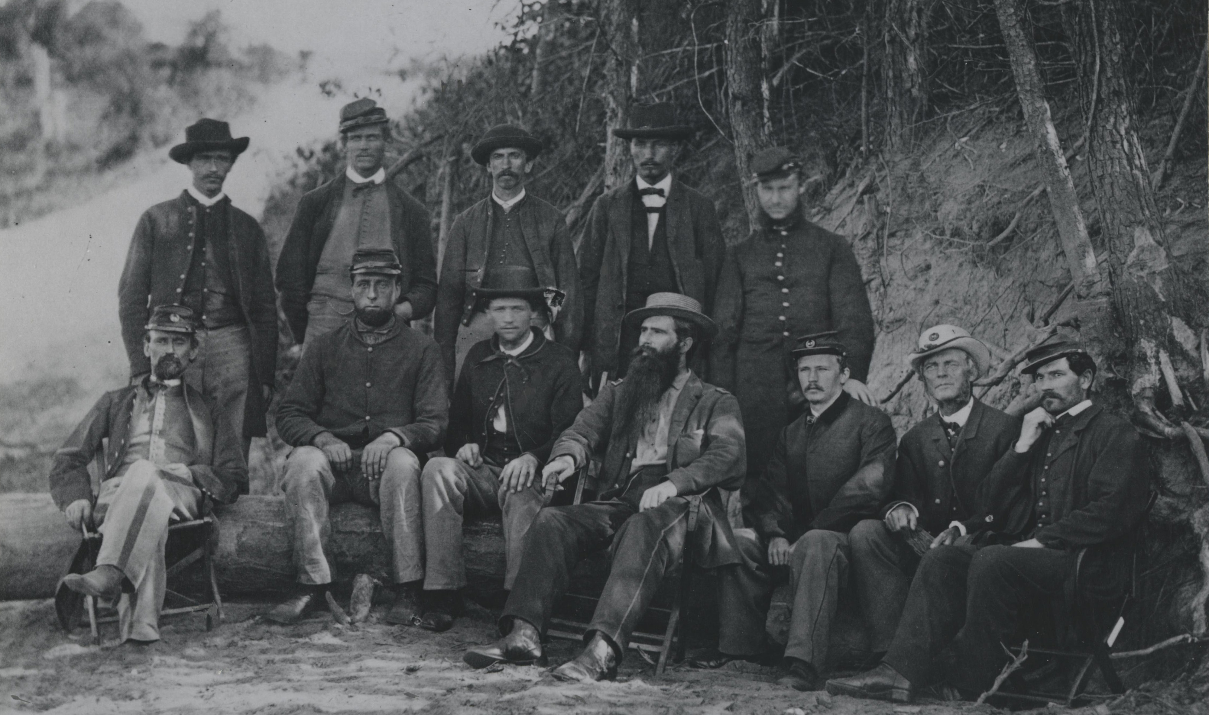
The advent of techniques in photomicrography in the 1800s unlocked an invaluable resource for scientific researchers. Lt. Col. Joseph Janvier Woodward, Assistant Surgeon General and head of the Medical and Microscopic Sections at the Army Medical Museum (now National Museum of Health and Medicine/NMHM), in partnership with the museum's Assistant Surgeon Maj. Dr. Edward Curtis, sought to improve upon these techniques, apply them to medical research, and harness findings for a deeper understanding of anatomy and disease than had yet been achieved.

Lt. Col. Joseph Janvier Woodward, Assistant Surgeon General and head of the Medical and Microscopic Sections at the Army Medical Museum (now National Museum of Health and Medicine) (NMHM, OHA 187, The Armed Forces Institute of Pathology: Its First Century, 1862-1962).
Photomicrography is the process of using a microscope to photograph a magnified image of microscopic specimens. In its simplest form, an image of a specimen viewed through a microscope is illuminated by a light source onto a screen or paper, then this projected image is photographed. Invented at the turn of the 19th century for the furtherment of scientific research, there are many theories on who developed the process. Some attribute it to Thomas Wedgwood and his partner Humphry Davy who captured vague images of objects on silver nitrate by using sunlight. However, it wasn't until the 1830s that scientist and photographer William Henry Fox Talbot captured the first photomicrographs: plant sections. Following this, photomicrography rose to fame in the scientific and medical community for its ability to document the anatomy of microscopic specimens.
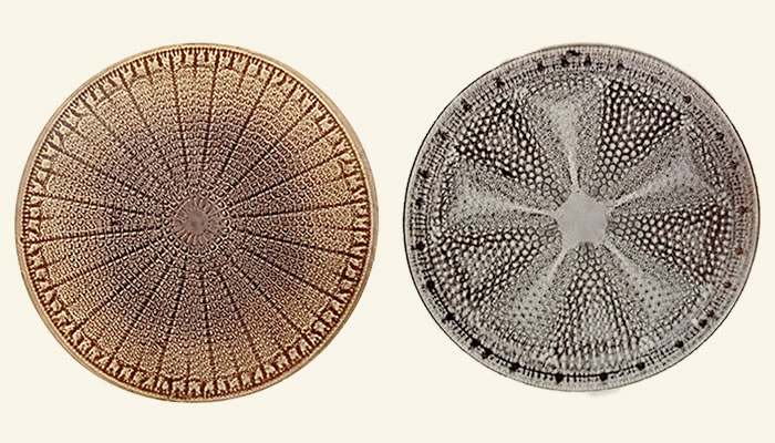
Photomicrograph of diatoms taken by Lt. Col. Joseph J. Woodward and from the NMHM Woodward Photomicrograph Collection (NMHM, OHA 79, No. 178 and 268).
Inspired by Talbot and other photomicrographic pioneers, in the 1860s Woodward headed the newly established Photographic Bureau for collections of photomicrographic material at the Army Medical Museum. Woodward experimented with the techniques of photomicrography, looking for innovations in the process and applications for medical research. Pioneering the use of aniline dyes in staining tissue, so that certain parts would become more visible under the microscope, Woodward, with assistance from Curtis, also developed the use of artificial lighting. This improved clarity and definition of the specimen's image when enlarged.
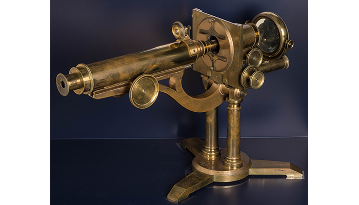
Zentmeyer's "Grand American Model" Compound Microscope, ca. 1862 (170614-D-MP902-002: Released/Courtesy of NMHM). Lt. Col. Joseph J. Woodward used this microscope for his pioneering work in photomicrography, which provided extraordinary insight into the physiological processes associated with various diseases before the development of microbiology and the germ theory of disease. This microscope and others belonging to Woodward are among those held in the Billings Microscope Collection at NMHM, the largest and most comprehensive collection of microscopes in the world.
Intrigued by the Virchow Archive at the Army Medical Museum, Woodward applied his innovative photomicrography techniques to experiment with and research disease. Rudolf Ludwig Carl Virchow, a German scientist known as the father of pathologic anatomy, began using microscopes to study disease, one of his greatest accomplishments noting that the whole organism does not get sick, only certain groups of cells.
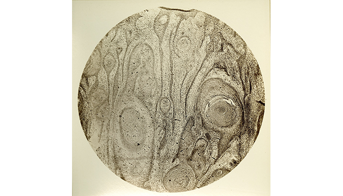
Photomicrograph of epithelial cancer of leg showing marrow, cancer cylinders, and "globules epidermique." Captured by Lt. Col. Joseph J. Woodward and from the NMHM Woodward Photomicrograph Collection (NMHM, OHA 79, No. 007).
Woodward was inspired by Virchow's use of microscopy to found cellular pathology, the study of microscopic anatomy of organs and tissues. Considering the microscopic images produced by Virchow, and with help from Curtis, Woodward took images of human and animal tissues, wool and linen fibers, and tiny unicellular plants. In one case, Woodward studied the nature of cancerous tumors, using photomicrography technology to see and document cancer cells in a tumor. Woodward eventually published his design improvements in photomicrography and his discoveries of clinical cases.
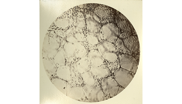
Photomicrograph of breast cancer showing the invasion of adipose tissue. Captured by Lt. Col. Joseph J. Woodward and from the NMHM Woodward Photomicrograph Collection (NMHM, OHA 79, No. 056).
Through Woodward's experiments, the Army Medical Museum became a center for photomicrographic use in medical cases. Woodward's documentation presented doctors and medical researchers with visual evidence of the progression of disease and patients' responses to treatment. The legacy of Woodward's work at the Army Medical Museum has inspired continued improvements in techniques of medical research for both civilian and military medicine, such as contemporary photomicrographic imaging using electron microscopes.
Today, NMHM has a microscope collection containing more than 1,700 items, more than 6,000 photomicrograph glass-plate negatives, and a large number of paper-printed photomicrographs; many were produced by Woodward himself.
Resources
Explore the Otis Historical Archives and some of NMHM's Photomicrograph Collections:
- Photomicrograph Collection (OHA 79) (PDF 446 KB)
- Woodward Collection (OHA 363) (PDF 466 KB)
- Arnold Photomicrographs (OHA 101) (PDF 415 KB)
- Benecke Photomicrographs (OHA 112) (PDF 439 KB)
- Giraud Photomicrograph Collection (OHA 173) (PDF 323 KB)
- Gray Photomicrograph Collection (OHA 178) (PDF 448 KB)
- Richmann Photomicrographs (OHA 289) (PDF 300 KB)
Relevant Links:
Woodward JJ, On the use of monochromatic sunlight as an aid to high-power definition:
https://collections.nlm.nih.gov/catalog/nlm:nlmuid-101214876-bk
Woodward JJ, The application of photography to micrometry, with special reference to the micrometry of blood in criminal cases:
https://collections.nlm.nih.gov/catalog/nlm:nlmuid-101309679-bk