An official website of the United States government
 ) or https:// means you've safely connected to the .mil website. Share sensitive information only on official, secure websites.


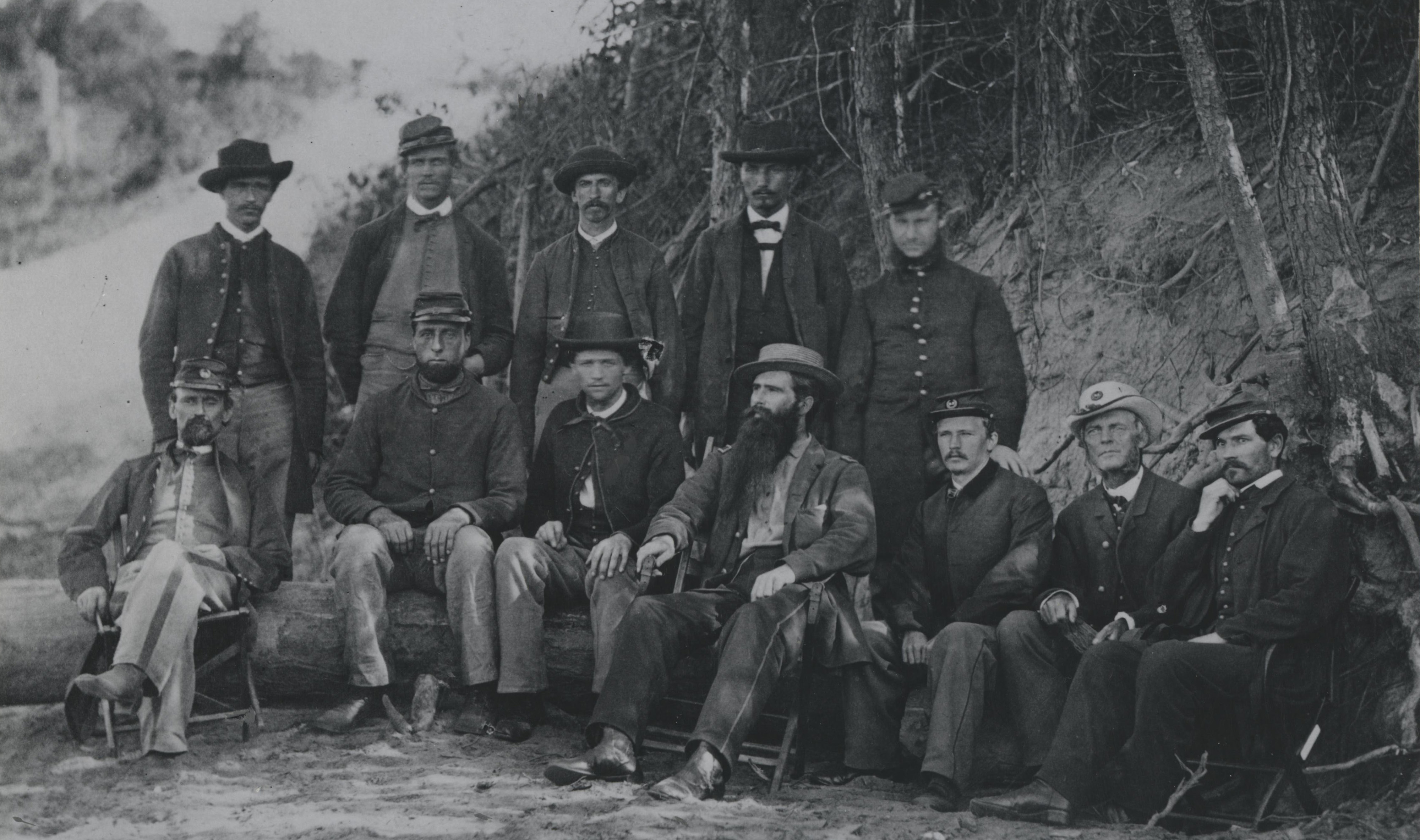
In 1874, the Army Medical Museum (now known as the National Museum of Health and Medicine) collected its first microscopes under curator Lt. Col. George A. Otis. These early additions to the collection mark the beginning of the Billings Microscope Collection, which today contains more than 1,200 microscopes ranging from 1650 to present-day, and is complimented by related materials present in the Otis Historical Archives.
While Lt. Col. John S. Billings toured Europe in search of historic medical items, he met John Mayall, Jr., a member of the Royal Microscopical Society in London, England. As a result of this relationship, the museum purchased 141 instruments from Mayall between 1884 and 1888, the most notable of these, "Hooke's model."
From this beginning, the Billings Microscope Collection has developed into one of the largest and most comprehensive collections of microscopes in the world, encompassing instruments which were used to identify the vector for yellow fever by museum curator and Army physician Maj. Walter Reed, and recent technologies like scanning electron microscopes and laser scanning microscopes.
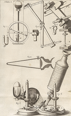
Drawing of the design and function of the Hooke microscope by Robert Hooke, and published in "Micrographia." (Image Courtesy of the National Library of Medicine)
"Hooke's microscope is generally admitted to be the editio princeps of the compound microscope, he being the inventor of that form," according to Billings in his written correspondence with the Army Medical Museum; accessible in the Otis Historical Archives. Hooke commissioned Christopher Cock to build this microscope, which shared several features with period telescopes including the eyecup and a separate tube for focusing. However, the microscope functioned oppositely to a telescope focusing on the enlargement of something small. Publishing his work in the first treatise on microscopy titled "Micrographia," Hooke recorded his observations including one of the most revolutionary discoveries in biologic and microscopic history: the discovery of "cells," a term he coined.
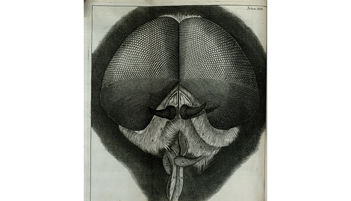
Close-up detail of the eyes and head of a grey drone fly viewed through the Hooke microscope, published in Robert Hooke's "Micrographia." (Image Courtesy of the National Library of Medicine)
Hooke's discovery from this microscope forced the realization of the existence of structures that were too small to be seen by the unaided eye. Antoni van Leeuwenhoek, one of the greatest early microbiologists–probably the first to observe microorganisms which he called animalcules and who discovered protozoa and bacteria–was directly inspired by Hooke's "Micrographia," according to Brian J. Ford, author of "The Leeuwenhoek Legacy." Ford claims that when Leeuwenhoek prepared his first package of specimens for the Royal Society in London, they were exactly the same sections of plant and animal material as those described by Hooke in his publication, and they were listed in the same order and in almost the same words.
Though innovations in microscopic technology have vastly improved, the modern era of biological and medical research stem from Hooke's microscopical investigation of matter. Today, microscopy is used to improve civilian and military life. For example, the United States Army Medical Research Unit-Kenya (USAMRU-K), established in 1973, helps to develop and test improved ways to treat infectious diseases that could threaten the U.S. military. Introducing thirty-six microscopy courses for lab technicians in African countries, USAMRU-K uses microscopy to combat malaria. Similarly, the Walter Reed Army Institute of Research used transmission electron microscopy to study the Zika virus and isolate the strain in a thin layer of specimen, revealing a clear image of the virus' structure. This information allowed Walter Reed's researchers to understand the disease better and establish trials for treatment in Nov. 2016.
"I have dealt somewhat fully with Hooke's microscopes, but not more fully than I think they deserve in view of the fact that he was the first to give a real impetus to English microscopy in the two main branches in which it is divided–in both of which his influence was conspicuous," stated Mayall in the "Cantor Lectures" of 1888.
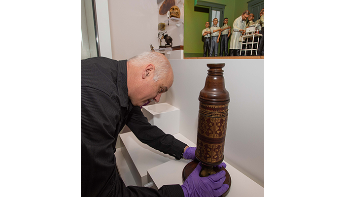
National Museum of Health and Medicine Historical Collections Manager Alan Hawk installs the Hooke microscope during the March 11, 2019 Exhibits Quarterly Review at the museum in Silver Spring, Maryland. (Disclosure: This image has been cropped to emphasize the subject.) (Department of Defense photo by Matthew Breitbart/ Released)
While the Hooke microscope has been off display for some time, it was recently reinstalled during NMHM's Exhibit Quarterly Review, a process when display cases are opened, exhibits are assessed, and preventive conservation is performed. The microscope replaced a corrosion cast of sea elephant lungs.
Resources
Ford, Brian J., The Leeuwenhoek Legacy, Biopress, Bristol, 1991
Relevant Links:
Improving Malaria Diagnosis in Africa, One Lab at a Time
https://www.army.mil/article/40343/improving_malaria_diagnosis_in_africa_one
_lab_at_a_time
Human Trials Begin for Army-Developed Zika Vaccine
https://dod.defense.gov/News/Article/Article/999584/human-trials-begin-for-army-developed-zika-vaccine/