An official website of the United States government
 ) or https:// means you've safely connected to the .mil website. Share sensitive information only on official, secure websites.


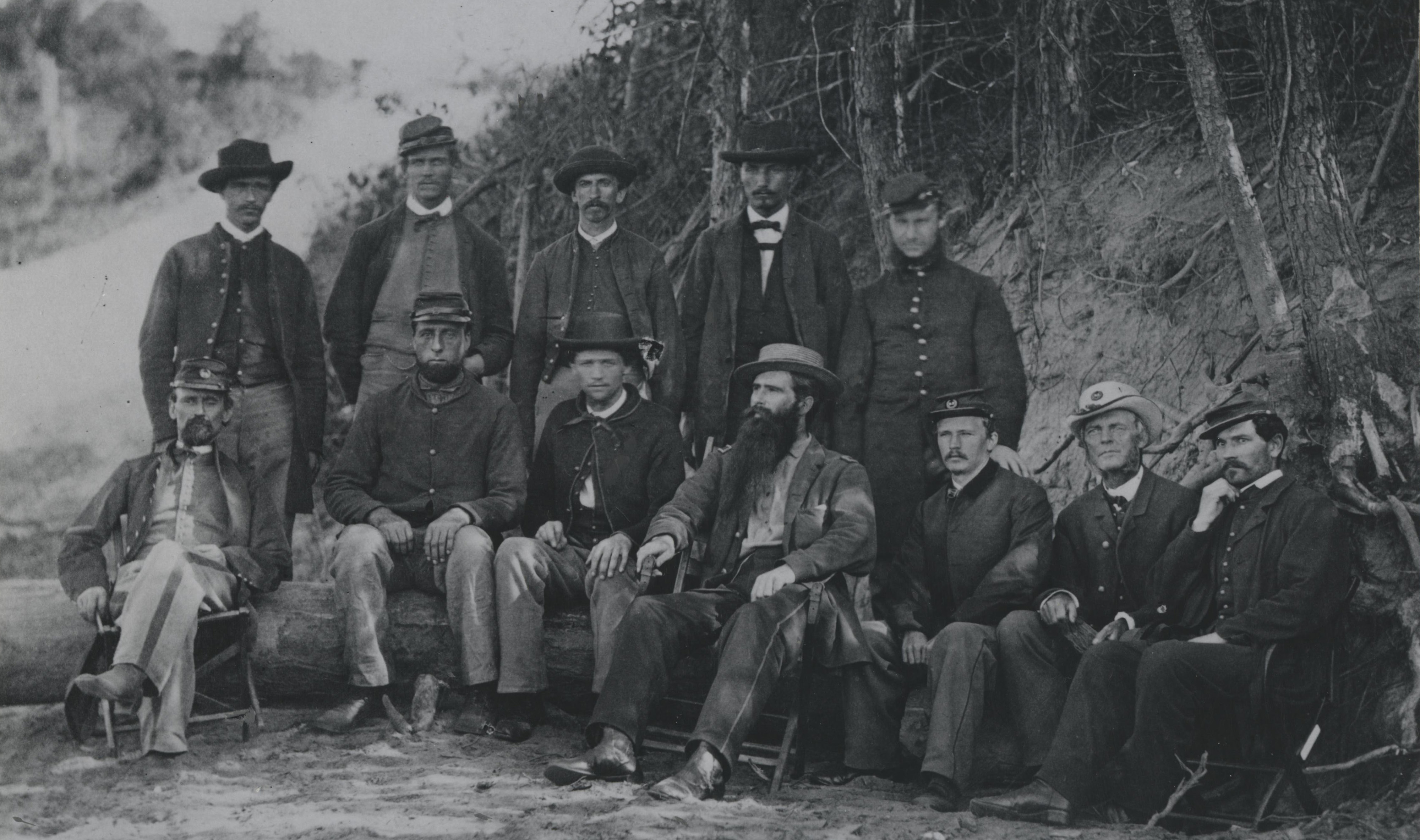
Our Anatomical Collections contain skeletal specimens that highlight the history of military and civilian medicine dating from the American Civil War and the founding of the museum in 1862. NMHM curates more than 6,400 skeletal specimens that display a variety of pathological conditions, including battlefield trauma, amputation, bacterial infection, and bone cancers and tumors.
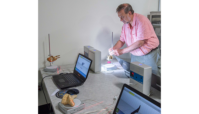
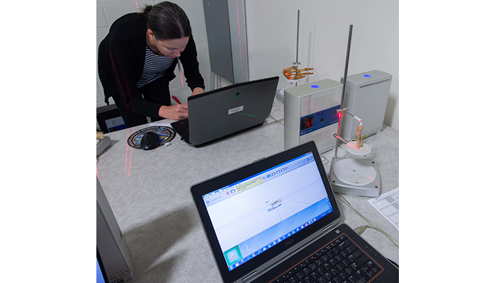
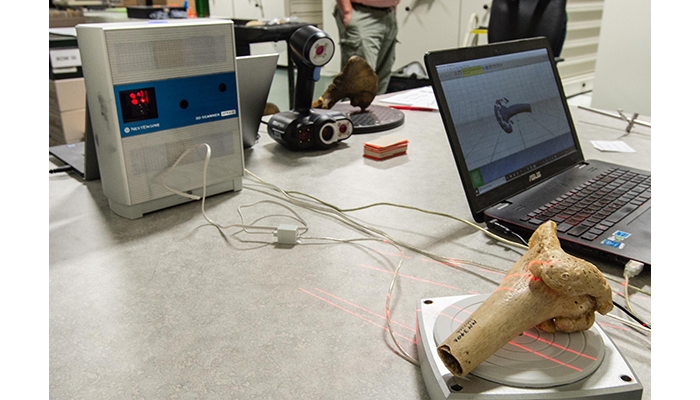
Virginia Commonwealth University Instructor of Anthropology Dr. Bernard Means prepares an anatomical specimen for a 3D scan at the National Museum of Health and Medicine at U.S. Army Garrison Fort Detrick-Forest Glen Annex in Silver Spring, Maryland in May 2019. (Department of Defense photo by Matthew Breitbart/ Released)
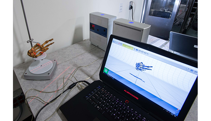
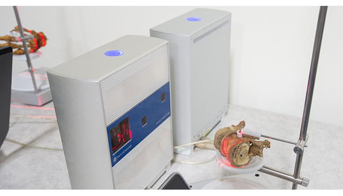
NMHM is collaborating with Virginia Commonwealth University's Virtual Curation Laboratory and Federal Bureau of Investigation's forensic anthropology laboratory to digitize and disseminate high-quality 3D models via an online portal, enabling scholars and educators to manipulate, analyze, and print the 3D models from anywhere in the world. These models represent unique traumatic injuries or manifestations of infectious disease that are otherwise unavailable for teaching or study without direct access to museum collections. Manipulation of the digital models allows researchers to study aspects of bone healing and response to disease in minute detail. The ability to download and print the 3D models allows students to interact with tactile and tangible examples of pathological conditions, rather than learn exclusively from pictures.
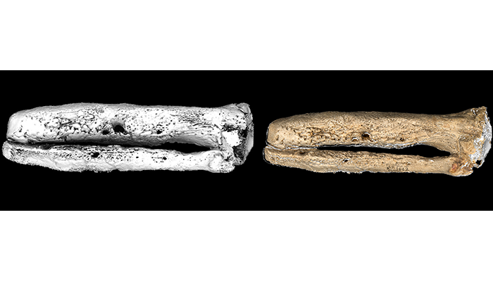
Figure 1 Digital model of left tibia/fibula of a 24-year-old male with osteomyelitis 19 months after primary amputation for gunshot injury. Civil War, 1864. Left: MicroCT. Right: MicroCT with partial photo wrap. (AFIP 1002692)
The 3D models are produced by scanning the specimens with sub-millimeter micro-computed tomography (micro-CT) to provide detailed images of internal and external structures. The models generated from micro-CT data (see figure 1 left) are enhanced with photorealistic capture of external surfaces through laser scanning and photogrammetry (see figure 1 right) and can provide specimen models for research, classroom interaction, and 3D printing that reflect the complex bony reactions of the actual specimen.
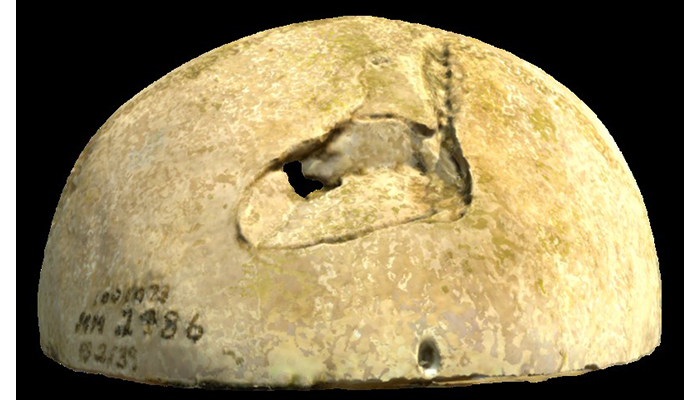
Figure 2 Digital model of the frontal bone of 22-year-old male with a depression fracture from conoidal musket ball. Civil War, 1863. Laser surface scan. (AFIP 1001073)
For models where visualization of the internal structures is not as critical, such as certain fractures, external lesions, or examples of human variation, external surface capture is being implemented with a laser scanner or structured light scanner (see figure 2). By combining imaging capture techniques, the same specimens can be made accessible on multiple web-based platforms to users with varying levels of experience or analytical needs.
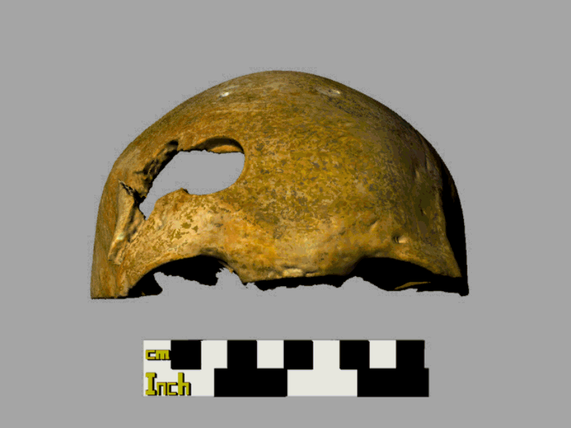
Figure 3 Digital image of the frontal bone of 24-year-old male with gunshot injury with circular trephination. Civil War, 1863. Animation from laser scanner. (AFIP 1001317)
Simpler representations, such as movie files or animated GIFs, can also be generated from the micro-CT slices, 3D volume renderings of the micro-CT data, and surface scans (see figure 3). This will allow the model to be viewed from all sides without additional manipulation.
Many institutions with courses in paleopathology, skeletal biology, and forensic anthropology do not have reference collections available for hands-on learning and do not have access to museum collections. Therefore a digital repository of specimens can provide an unprecedented and unique resource for researchers and educators. The sharing of these military medical assets will improve historical knowledge and diagnostic capabilities in the fields of medicine and anthropology. Access to the full data sets for the scanned specimens are available by request through the museum.
Acknowledgement: Digital models by VCU's Virtual Curation Laboratory.
Resources
Virtual Curation Laboratory Blog
https://vcuarchaeology3d.wordpress.com/
FBI's Handbook of Forensic Services
https://www.fbi.gov/file-repository/handbook-of-forensic-services-pdf.pdf
Relevant Links:
NMHM Presentation at 2019 Paleopathology Association Meeting
Lab 3D Scans Human Skeletal Remains Dating Back to the Civil War
https://news.vcu.edu/article/Lab_3D_scans_human_skeletal_remains_dating_back_to_the_Civil