An official website of the United States government
 ) or https:// means you've safely connected to the .mil website. Share sensitive information only on official, secure websites.


In the years after the Civil War, the Army Medical Museum (predecessor to the National Museum of Health and Medicine) began purchasing large numbers of anatomical preparations to expand the scope of the museum's collections. By 1867, multiple specimens were purchased from the premier anatomical specimen preparators in Paris, France such as Pierre Vasseur and Louis Auzoux, for a newly-developed Anatomical Section to complement the existing Pathological Section, comprised at that time of trauma specimens from the Civil War. A few of the specimens, including a full skeleton of a 25-year-old French male, were mounted using the Beauchêne method. Due to this unique preparation, the Beauchêne specimens became rare and valuable assets to the museum's teaching collections.
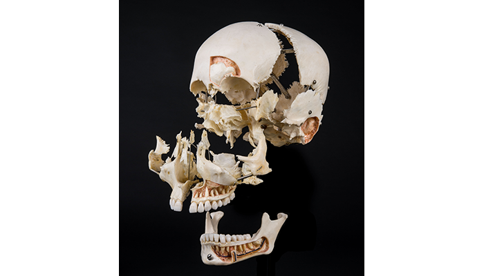
A skull prepared in the Beauchêne method, also known as an "exploded" specimen, shows individual bones, sinuses and cutaway views of the nerves and blood vessels related to the teeth. (150326-A-MP902-077: NMHM photo)
Anatomist and surgeon Edmé François Chauvot de Beauchêne is the namesake of the technique. Also called the exploding skeletal technique, the Beauchêne method relies on the deconstruction or disarticulation of the bones and joints. The separated bones are spaced apart and then mounted in anatomical position, often with wires. This detailed and intricate process was often time-consuming and labor intensive.
Milton Hildebrand's Anatomical Preparations describes the long process:
"Clean the skull meticulously, bleaching it if desired, and degreasing it if necessary. The skull is then boiled or macerated until the cranial sutures are loosened. At this point the skull bones can be disarticulated by hand while the skull is wet. If the cranial bones are difficult to separate, the braincase should be further soaked until disarticulation is possible. Once the bones are disarticulated, they are dried and mounted according to the desired design of the creator. Wires or small strips of celluloid are used to space the bones, and individual bones or assembled units of the skull are supported on a wooden base by heavy wires."
Popularized for use in anatomical study in the mid-19th century, this innovative technique reveals the structure of the specimen and the relationships between bones. The preparation allows the viewer to understand the specimen through its individual parts, as well as how the pieces fit together collectively as a whole.
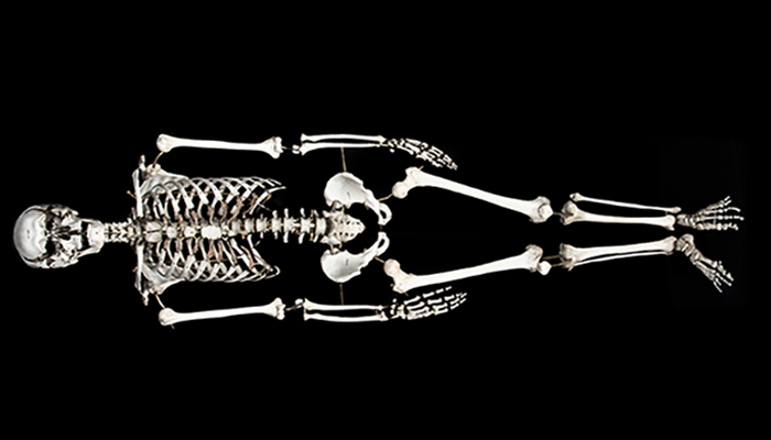
Specimen No. 2, a Beauchêne prepared skeleton, was purchased by Bassange of Paris from Auzoux in 1867. The skeleton is on display at the museum. (120907-A-MP902-001: NMHM photo)
When Surgeon General Joseph K. Barnes ordered the purchase of these specimens, he did so hoping they would enhance the teaching collections of the museum. Previously, museum accessions focused on injury and disease; however, the scope of the museum grew as "specimens of [normal] human anatomy were soon found not only useful but necessary additions to the original collection," said former curator John Shaw Billings. These specimens demonstrated an understanding of normal anatomy and could be used to compare against specimens of a pathological nature.
Beauchêne preparations continue to hold value within our collections as it "is the best method for visualizing the interrelations of all of the skeletal components of the head simultaneously. The articulations of many individual bones with one another can be obscured in a normal complete human skull. In particular, the relative positions of the sphenoid, vomer, and ethmoid bones are more clearly visualized," said Anatomical Curator Brian Spatola.
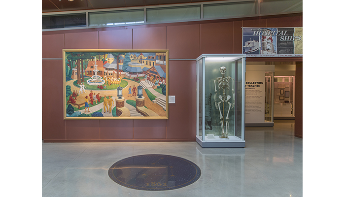
The lobby of the museum exhibits a 1944 painting of Forest Glen by Jack McMillen, and Specimen No. 2 of the Anatomical Collections: a Beauchêne prepared skeleton. (200117-D-TY520-0001)
Today, Beauchêne prepared human specimens are harder to find and rarely available as technology has made it easier to "explode" skeletons with plastic models and 3D animation. However, the museum continues to display its Beauchêne preparations. The museum has five Beauchêne preparations available to visitors and researchers: one full skeleton, three human crania, and one non-human cranium (a primate).
Resources
Hildebrand, Milton. Anatomical Preparations. University of California Press, 1968.
Lamb, D.S. A History of the United States Army Medical Museum, 1862 to 1917. Washington, 1917.
http://resource.nlm.nih.gov/12710920R
Spinner, Robert J., Vincent, Jean-François, Wolanskyj, Alexandra P. "Discovering the Elusive Beauchêne: The Originator of the Disarticulate Anatomic Technique." Clinical Anatomy 24, no. 7 (October 2011): 797-801.
https://www.ncbi.nlm.nih.gov/pubmed/21898884
Relevant Links:
Checklist of Preparations and Objects in the Section of Human Anatomy of the United States Army Medical Museum
https://collections.nlm.nih.gov/catalog/nlm:nlmuid-101589481-bk
Retracing the Observations and Footsteps of Beauchêne Fils: A Blueprint for Scientific Research
https://www.ncbi.nlm.nih.gov/pubmed/30117198