An official website of the United States government
 ) or https:// means you've safely connected to the .mil website. Share sensitive information only on official, secure websites.


Marie Francois Xavier Bichat (November 14, 1771 to July 22, 1802), a French anatomist and physiologist, is best remembered as the father of modern histology and descriptive anatomy. Despite working without a microscope, Bichat was the first to introduce the notion of tissues as distinct entities and maintained that diseases attached to tissue rather than whole organs.
Bichat's observations helped found the branch of science called histology, the study of tissue, which led to other similar avenues of study like histopathology, the study of diseased tissue.
Finished routine Haematoxylin and Eosin (H&E) stained slide preparations readied for microscopic examination. (NMHM photo: 210407-D-D0458-0001)
Histotechnicians make microscopic slide preparations of human, animal, and plant tissue for microscopic diagnosis and analysis. In a hospital or clinical setting, each tissue specimen submitted from the operating room is routinely sent to a histology lab for preparation to rule out or confirm disease.
A histology lab may specialize in one or many areas of anatomy, paleohistology (fossil study), or even the study of plant tissues, and may operate differently based on the specific line of study.
As a histotechnician on staff at the National Museum of Health and Medicine, I have the responsibility to care for the slide-based collections. My experience has been shaped by work in a hospital setting as well as at the Armed Forces Institute of Pathology, where I worked in the histology lab until its closure in 2011. When I transitioned to NMHM, I brought my skills of a medical laboratory professional, which I now use when I conserve the museum's slides and wet tissue specimens. From my personal experience, below are the descriptions of 10 basic stations in the histology lab. Equipment is spaced and arranged in the order the specimen goes through the preparatory process from start to finish to increase efficiency.
- Specimen entry: The specimen is delivered to the lab in a fixative (a substance used to preserve and set), usually a formalin fixative. In a clinical setting, such as a hospital, the specimens are routinely received from the operating room in a container of 10% formalin solution. Large specimens, such as a complete organ (e.g., a lung or kidney), may be received fresh without a fixative until after gross examination (review of the organ with the bare eye) and dissection. If needed, a fixative is added at entry. From this point, the specimen is given an accession number for identification. This ID number is used to record the information accumulated from the microscopic examination of the specimen and is kept on record for reference. Non-human specimens are also accessioned with ID numbers and filed for future reference and study. The different symbols used as ID numbers vary from lab to lab as each has their own system for recording cases.
- Grossing: This station reports the received specimen's appearance and measurements while noting any remarkable characteristics. The specimen is also dissected and submitted into cassettes for processing.
- Processing: The specimens go through fixation, dehydration, clearing, and wax infiltration; this will remove all the water from the specimen and replace it with wax to make it easy to handle and cut into slides. This is routinely done on an automated tissue processor.
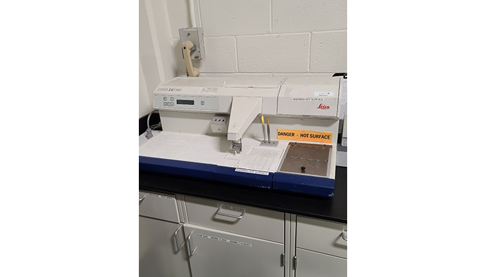
Embedding station equipment at the National Museum of Health and Medicine. This machine dispenses molten paraffin wax (heated between 56 and 60 degrees Fahrenheit). (NMHM photo: 210406-D-D0458-0001)
- Embedding: The tissue is placed in a metal mold surface with paraffin wax, and a plastic cassette is placed on top of the mold. Both together are placed on a refrigerated plate to create a solid paraffin tissue block. This is the preparatory stage for microtomy.
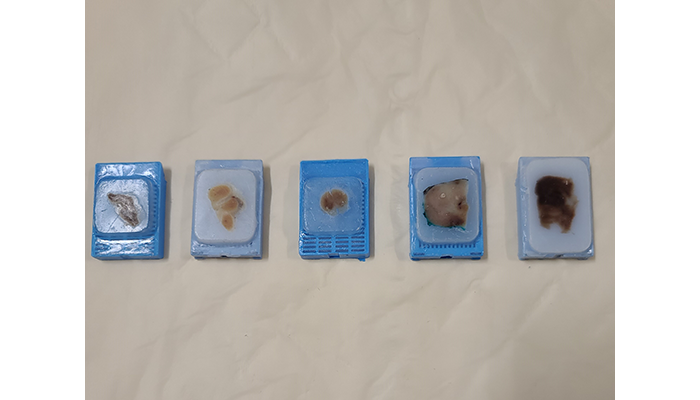
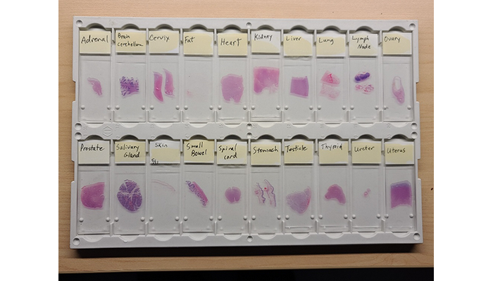
Tissues embedded in paraffin wax in preparation for microtomy stage. Specimen identification from left to right: adrenal gland, brain cerebrum, brain stem, prostate, heart. (NMHM photo: 210407-D-D0458-0001)
- Microtomy: The tissue embedded in the wax is sectioned with a microtome (a cutting tool) into thin slices to be placed on a microscope slide in preparation of the staining process.
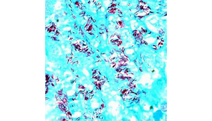
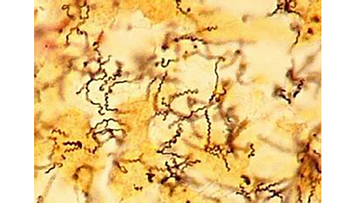
- Staining: The slide preparations are submitted to routine H&E (haematoxylin and eosin) staining. Additional stain protocols involve special stains and/or immunohistochemical staining (a process that uses antibodies to detect the location of proteins and other antigens in tissue sections) dependent on what the pathologist suspects or needs to rule out in their microscopic diagnosis.
Photo Gallery:
- Coverbonding: The process of placing a "glass" cover over the tissue on the prepared slide to maintain the "flatness" of the tissue and preserve it for future viewing is coverbonding. This takes place immediately after each slide has been stained.
- Labeling: Each specimen carries the accession number given at specimen entry. The slide preparation has two labels: one is on the slide (written in marker or diamond pen) and the second is a handwritten or printed label placed on top of the written number. The printed label creates an easy visual ID of the slide, and the number on the slide itself is more permanent if the printed label is removed from the slide.
- Delivery: All slides are placed in slide folders in the numerical order of the day and delivered to the pathologists with the patient information and reports. The pathologist is now equipped to provide the microscopic diagnosis of each case and/or request additional preparations in the form of recuts, serial sectioning, special stains, or immunohistochemistries.
- Filing: The filing of blocks happens immediately after the slides are delivered to the pathologist. All blocks are filed by year and numerical order for efficient retrieval. All slides are filed after they are returned to the histology lab and after the coverbonding medium has dried.
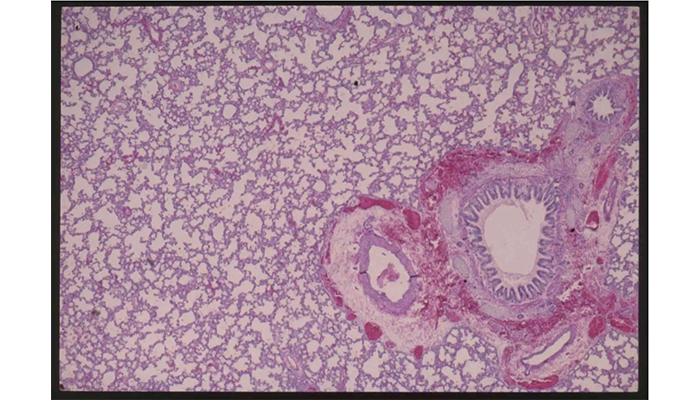
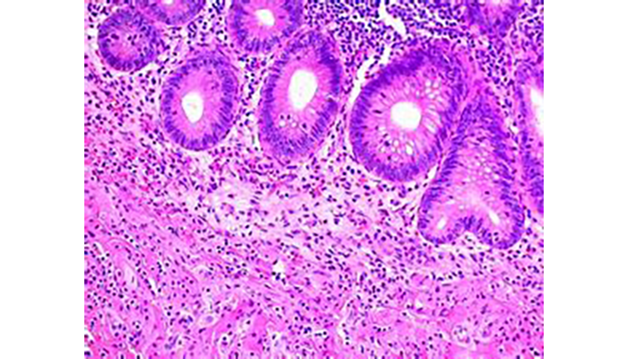
Histological slide from the National Museum of Health and Medicine of interstitial pneumonia, also called parvo virus, in a dog. (CD00064)
The museum does not create microscopic slide preparations. Instead we maintain a collection of histological slides, microscopes, microtomes, and the equipment and tools of the histologist, microscopist, and other medical laboratory professionals. Our slide collections are simply records of previous slides made for the purpose of microscopic examination and diagnoses. In the clinical field, microscopic slide preparations allow the pathologist to examine the tissue and confirm or rule out diseases. In the field of research, these same slides may be used for study, as in teaching or learning more about diseases. The museum's collections hold slide preparations of every type of human tissue from every part of the human body; all body organs are represented.