
Visibly Human Health and Disease in the Human Body
The Cardiovascular System
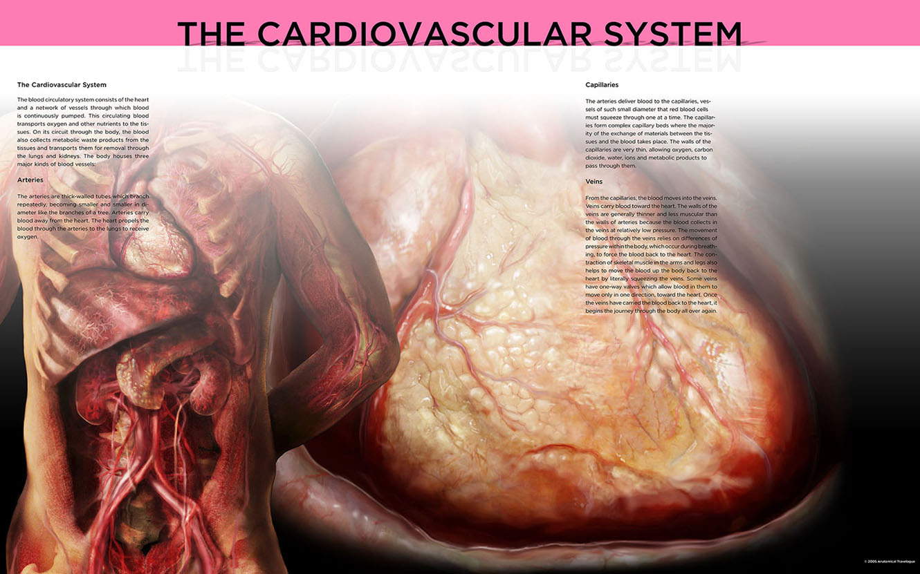 The blood circulatory system consists of the heart and a network of vessels through which blood is continuously pumped. This circulating blood transports oxygen and other nutrients to the tissues. On its circuit through the body, the blood also collects metabolic waste products from the tissues and transports them for removal through the lungs and kidneys. The body houses three major kinds of blood vessels:
Veins
From the capillaries, the blood moves into the veins. Veins carry blood toward the heart. The walls of the veins are generally thinner and less muscular than the walls of arteries because the blood collects in the veins at relatively low pressure. The movement of blood through the veins relies on differences of pressure within the body, which occur during breathing, to force the blood back to the heart. The contraction of skeletal muscle in the arms and legs also helps to move the blood up the body back to the heart by literally squeezing the veins. Some veins have one-way valves which allow blood in them to move only in one direction, toward the heart. Once the veins have carried the blood back to the heart, it begins the journey through the body all over again.
Arteries
The arteries are thick-walled tubes which branch repeatedly, becoming smaller and smaller in diameter like the branches of a tree. Arteries carry blood away from the heart. The heart propels the blood through the arteries to the lungs to receive oxygen.
Capillaries
The arteries deliver blood to the capillaries, vessels of such small diameter that red blood cells must squeeze through one at a time. The capillaries form complex capillary beds where the majority of the exchange of materials between the tissues and the blood takes place. The walls of the capillaries are very thin, allowing oxygen, carbon dioxide, water, ions and metabolic products to pass through them.
Healthy heart, plastinated
The human heart is a fist-sized, hollow, muscular pump which beats continuously, moving blood through the body. The human heart has four chambers. The two smaller, upper chambers are the atria (singular, atrium). The two larger, lower chambers are the ventricles. Both the atria and the ventricles have strong, thick walls primarily made up of myocardium: heart muscle. The chambers are separated from one another by one-way valves which allow the blood to flow in one direction only. The right side of the heart pumps blood to the lungs maintaining pulmonary circulation (Latin pulmo = lung). The left side of the heart moves blood throughout the remainder of the body, maintaining systemic circulation. This plastinated heart has been injected with dyes to show arteries and veins. Arteries are red. Veins are blue. 1987.3005.13 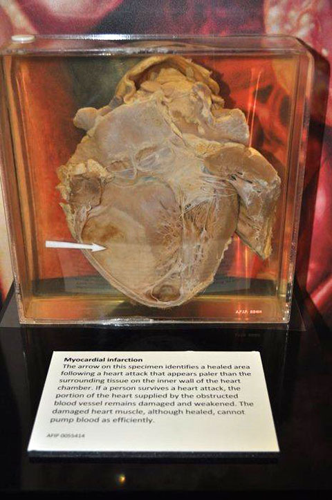
Myocardial infarction
The arrow on this specimen identifies a healed area following a heart attack that appears paler than the surrounding tissue on the inner wall of the heart chamber. If a person survives a heart attack, the portion of the heart supplied by the obstructed blood vessel remains damaged and weakened. The damaged heart muscle, although healed, cannot pump blood as efficiently. AFIP 0055414 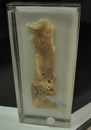
Gunshot wound of aorta
This normal aorta has a smooth inner surface. The arrow points to the tear caused by a gunshot wound. AFIP 1232818 |
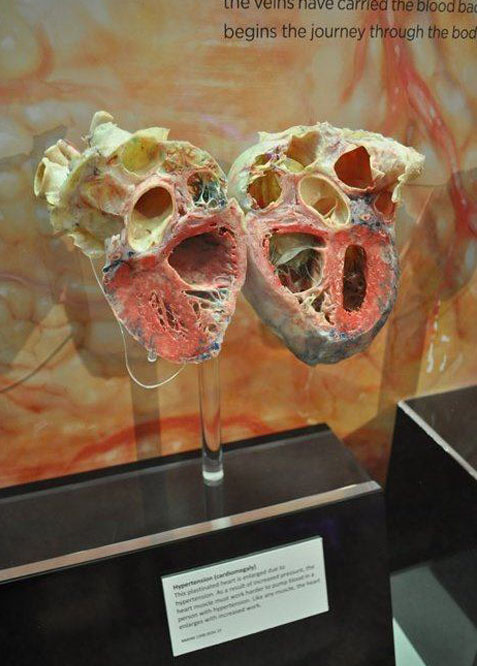
Hypertension (cardiomegaly)
This plastinated heart is enlarged due to hypertension. As a result of increased pressure, the heart muscle must work harder to pump blood in a person with hypertension. Like any muscle, the heart enlarges with increased work. NMHM 1998.0034.19 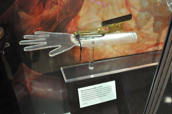
Marey sphygomograph, ca. 1870
This device produced a permanent record of the strength and rhythm of the pulse. The motion of blood pulsing through an artery in the wrist was transmitted by the spring to the stylus which recorded that motion on smoked paper. 1991.0053 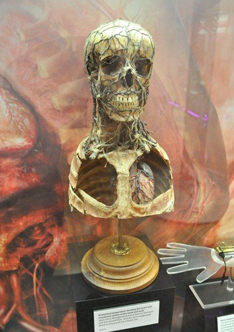
Anatomical preparation showing the heart in anatomical relation to the circulatory system of the head and neck.
Veins carry deoxygenated blood back to the heart and are colored blue. Arteries carry oxygenated blood to tissue and are colored red. MM4260 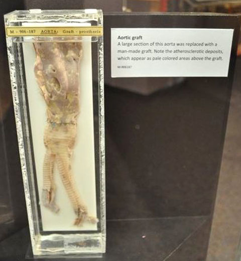
Aortic graft
A large section of this aorta was replaced with a man-made graft. Note the atherosclerotic deposits, which appear as pale colored areas above the graft. M-906187 |
- Visibly Human Health and Disease in the Human Body
- The Cardiovascular System
- The Urinary System
- Respiratory System
- The Lymphatic System
- The Musculoskeletal System
- The Liver and Hepatic System
- The Digestive System
- The Brain and Nervous System
- Psychiatric Patients at Forest Glen
- Skeleton of Spanish American War Veteran Showing Evidence of Severe Arthritis