
Visibly Human Health and Disease in the Human Body
Respiratory System
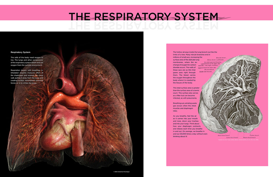 The primary function of the respiratory system is gas exchange. The lungs maintain a concentration gradient between oxygen and carbon dioxide, such that incoming oxygen-rich air is exchanged for outgoing air with high concentrations of carbon dioxide. The body requires oxygen in order to produce energy required for cellular activity.
Respiration begins with inhalation that occurs when the diaphragm, a dome shaped muscle below the lungs, is contracted. This contraction increases the negative pressure in the thoracic cavity pulling air toward the lungs. Air flows into the trachea (windpipe) that branches into the right and left bronchi and then flows further into various smaller bronchioles, which branch out like the limbs of a tree. Eventually air arrives at the end of the branches that terminate in millions of small sacs or alveoli where the exchange between oxygen and carbon dioxide occurs. The membranes of the alveoli are thin enough for gases to pass through them into the blood that is then carried to various tissues throughout the body.
This total surface area of adult human alveoli measures 70 m2 on average. This great amount of surface area makes it susceptible to infection by invading organisms; however, lung tissue is filled with macrophages that help to rid the body of foreign particles including pathogens. An infection of the lungs is called pneumonia.
After inhalation the diaphragm is relaxed and waste carbon dioxide gas is exhaled into the atmosphere. On average, we breathe in and out 20,000 times a day without even thinking about it!
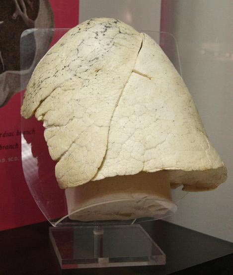
Healthy lung, left (2-lobes), air-dried
.
1998.0033.24 |
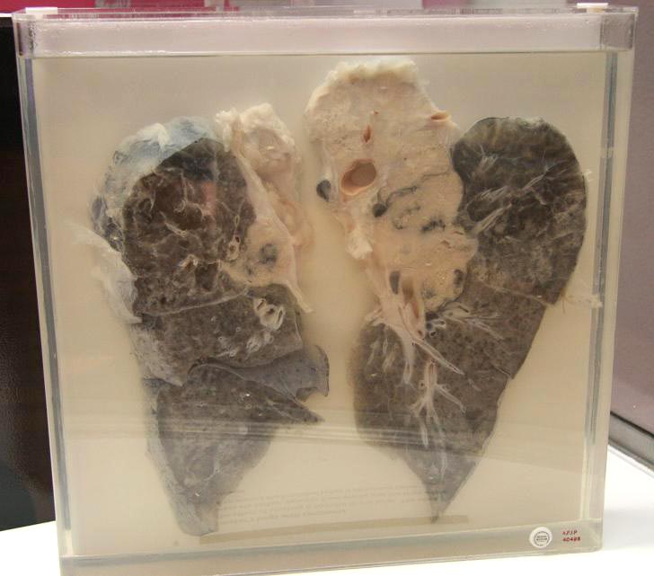
Smoker’s lungs with carcinoma
The effect of smoking is harmful to the lungs. The black areas are carbon deposits from smoke and the large white area around the bronchial tubes is cancerous tissue. AFIP 40498 |
|
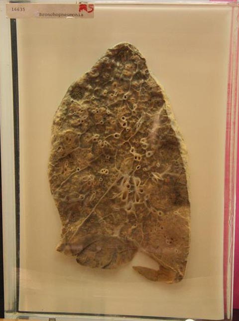
Infected lung with bronchopneumonia as a result of effects from influenza virus.
Camp Pike, Arkansas, 1918. The patient was admitted on October 10, 1918 with general aching, chills, fever, cough and a diagnosis of acute bronchitis and infection with influenza virus. Death occurred on October 25 due to bronchopneumonia. Bronchopneumonia often developed after infection with the influenza virus during the pandemic of 1918. This section of right lung shows multiple tan-colored nodules of consolidated inflammatory cells centered on the small airways called bronchioles. The body’s reaction to the influenza virus damaged the linings of the airways, making them vulnerable to bacterial infection. Today this could be effectively treated with antibiotics which were not available in 1918. AFIP 16635 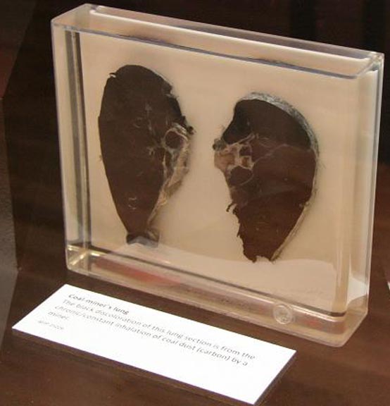
Coal-miner’s lung
The black discoloration of this lung section is from the chronic/constant inhalation of coal dust (carbon) by a miner. AFIP 25226 |
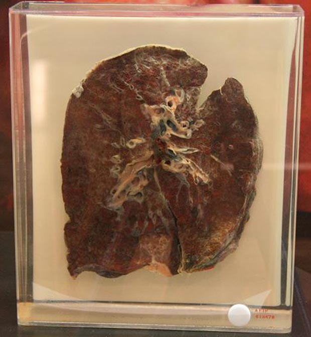
Iron-miner’s lung
This lung section is from a 76-year-old man who spent most of his working life mining iron ore. The black pigment is hematite. AFIP 613478 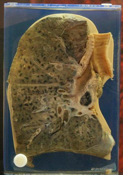
City dweller’s lung
Atmospheric pollutants inhaled during normal respiration can cause abnormal chemical deposits in the lungs. The deposits in this lung are black. This city dweller’s lung shows the extent of air pollution in the city. AFIP 86202 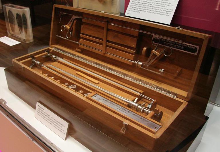
Bronchoscope
Lungs can be examined with x-rays and scans. Breathing sounds detected with a stethoscope can indicate the health of the lung. Devices like this bronchoscope can be inserted into the airways to see their interior. M-01300114 |
- Visibly Human Health and Disease in the Human Body
- The Cardiovascular System
- The Urinary System
- Respiratory System
- The Lymphatic System
- The Musculoskeletal System
- The Liver and Hepatic System
- The Digestive System
- The Brain and Nervous System
- Psychiatric Patients at Forest Glen
- Skeleton of Spanish American War Veteran Showing Evidence of Severe Arthritis