An official website of the United States government
 ) or https:// means you've safely connected to the .mil website. Share sensitive information only on official, secure websites.


On display at the National Museum of Health and Medicine is a hip bone with a Civil War bullet wound and a story of unusual medical intervention. NMHM staff wanted to learn more about the injury, beyond the text of the medical records, and carried out a micro-computed tomography scan of the hip bone which revealed new evidence that may hint at why infection of the wound persisted.
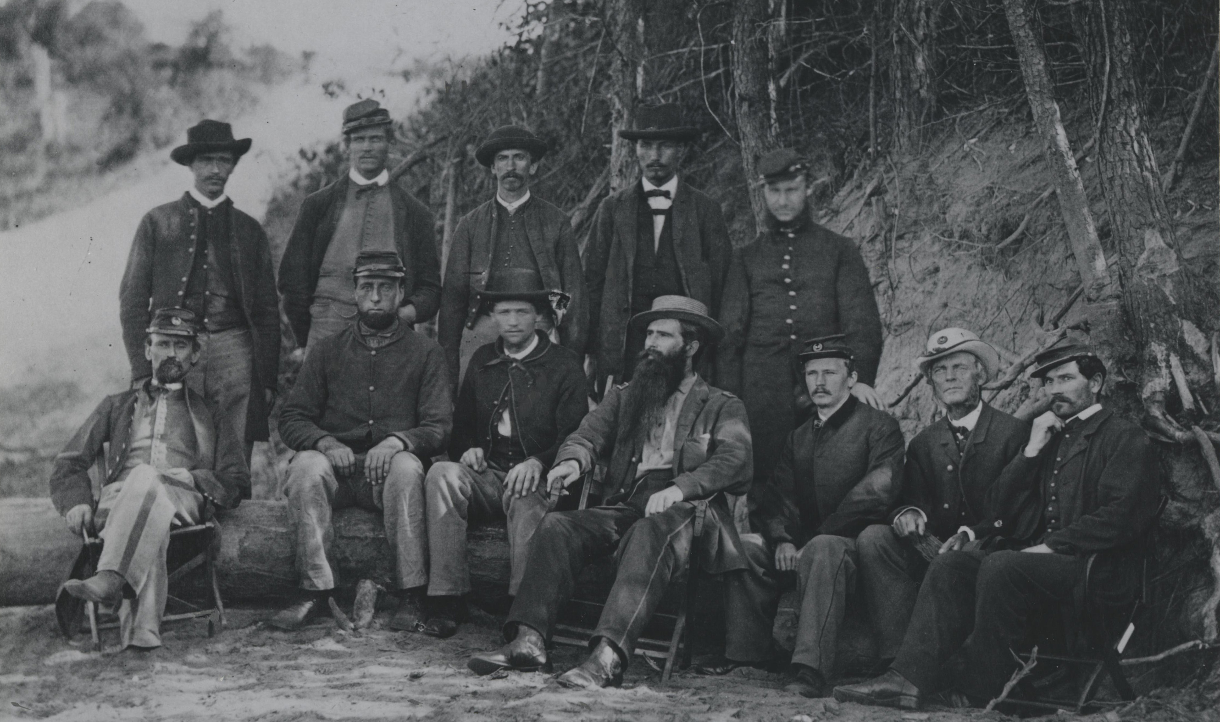
On July 1, 1862, Major Henry Alanson Barnum, 12th New York Infantry, was wounded at Malvern Hill, Virginia by a conoidal musket ball through the left hip. The bullet entered below and to the left of the navel, passed through the middle of the ilium, or hip bone, and emerged posteriorly, leaving a track from front to back of Barnum's torso.
The wound was regarded as fatal, and Barnum was left behind in a field hospital when Union forces withdrew to Harrison's Landing. On July 2, still alive, Barnum was captured by the Confederate army and taken to Libby Prison in Richmond, Virginia, eighteen miles away. More than two weeks later, he was returned to Union forces in a prisoner exchange and sent by boat to New York to rest and recover.
In October 1862, civilian doctor Alden March removed several fragments of dead bone from the infected injury. By January 1863, Barnum was recovered enough to return to the field as a colonel with command of the 149th New York Infantry. However, over the next several months, a large abscess formed and discharged around the wound. Dr. March opened the wound again but extracted no new fragments of bone. Barnum resumed his duties with the Union army and commanded his regiment at the Battle of Gettysburg in July 1863. He was injured two more times: in the arm at the Battle of Lookout Mountain near Chattanooga, Tennessee in November 1863 and in the right side at Peach Tree Creek, Georgia in 1864. Barnum ultimately received the brevet or honorary rank of major general in 1865.
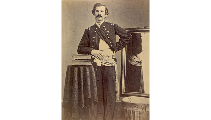
Henry A. Barnum. Photographed at the Army Medical Museum, August 1865. National Museum of Health and Medicine, Otis Historical Archives. (SP 93)
By January 1864, Barnum was emaciated, with pus regularly discharging from the hip. He returned to New York and was treated by renowned orthopedic surgeon Lewis Albert Sayre, who inserted an oakum, or tarred fiber, string saturated with Peru Balsam oil through the injury to establish a complete drainage from front to back. Several weeks later, Surgeon H.K. Hogan, U.S. Volunteers, replaced the oakum string with a candle wick which was gradually reduced in size.

Henry A. Barnum, ca. 1881. Henry A. Barnum Military Pension Records, certificate 78753, National Archives and Records Administration, Washington, DC. (NCP 003787)
In 1874, Dr. Sayre enlarged the wound to remove more bone, straightened the drainage canal, and inserted an India-rubber drainage tube, a process the doctor described in his 1884 medical casebook. A second tube was added in 1887. Barnum continued to wear both rubber tubes until his death from pneumonia on January 29, 1892.
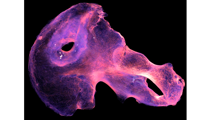
Digital model of left innominate of Maj. Gen. Henry A. Barnum. The periwinkle-colored ring represents dense reactive bone around the gunshot perforation. The white specks represent metal fragments embedded in the new bone. Micro-CT. (AFIP 1002020)
Although multiple surgeons and physicians attempted to resolve Barnum's injury over the course of thirty years, they were never successful in treating the chronic infection. A recent micro-computed tomography scan of Barnum's hip bone illuminated the hidden cause of infection: contamination from lead bullet fragments which contributed to the decades-long case of osteomyelitis, a bone infection.
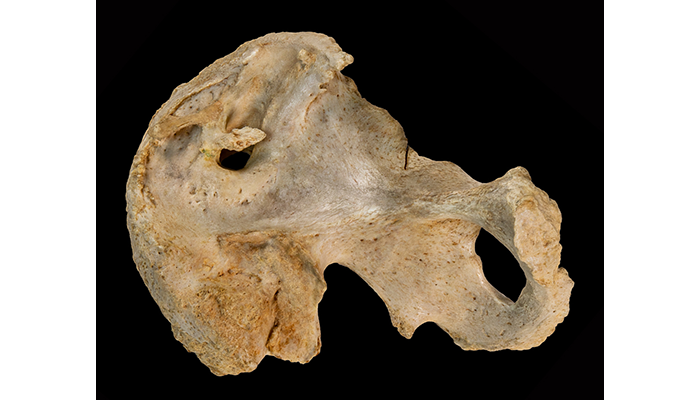
The left innominate of Maj. Gen. Henry A. Barnum. Barnum was injured at Malvern Hill, Virginia on July 1, 1862. The innominate exhibits osteomyelitis and sclerotic bone formation. (AFIP 1002020)
The bullet debris is obscured by dense reactive bone around the gunshot injury and is not visible to the naked eye, but is clearly seen in the CT scan. Scanning skeletal specimens like Barnum's hip bone helps the NMHM improve historical knowledge and understand individual post-traumatic experiences from the Civil War.
Resources
National Museum of Health and Medicine. AFIP 1002020, OHA 31, Otis Historical Archives.
Pearlstein, Kristen M., Terrie Simmons-Ehrhardt, Bernard K. Means, Brian F. Spatola, Angi M. Christensen, Richard M. Thomas, and Mary R. Mani. "Using 3D Modelling to Tell Individual Stories from the American Civil War." The 89th Annual Meeting of the American Association of Physical Anthropologists. April, 16, 2020.
Zampini, Jay M. & Henry H. Sherk. "Lewis A Sayre: The First Professor of Orthopaedic Surgery in America." Clinical Orthopaedics and Related Research 466, no. 9 (Sept. 2008): 2263-2267.
Relevant Links:
Modernizing Medical Museums Through the 3D Digitization of Pathological Specimens